CPQ Orthopaedics (2019) 3:1Review Article
A Review of Current Management of Distal Tibia Extraarticular
Fractures
Sujan Raj Paudel1*, Ankit Shrivastava2 & Ram Kewal Shah3
1Orthopaedic Surgeon, Scheer Memorial Adventist Hospital, Banepa, Nepal
2Orthopaedic surgeon, Janakpur Trauma Hospital, Janakpur, Nepal
3Head of Department, Department of Orthopaedics, Janaki Medical College, Janakpurdham, Nepal
*Correspondence to: Dr. Sujan Raj Paudel, Orthopaedic Surgeon, Scheer Memorial Adventist Hospital,
Banepa, Nepal.
Copyright © 2019 Dr. Sujan Raj Paudel, et al. This is an open access article distributed under the Creative Commons Attribution License, which permits unrestricted use, distribution, and reproduction in any medium, provided the original work is properly cited.
Received: 18 March 2019
Published: 01 April 2019
Keywords: Extraarticular Tibia Fractures; Distal Tibia Fractures; Nonunion of Tibia
Abstract
The treatment of distal tibia fractures is challenging. Various methods of treatment including
plaster, traction, splints, external fixation and internal fixation methods like intramedullary nails or
plates have been used to treat such fractures. The optimal treatment of unstable distal tibia fractures
without articular involvement remains controversial. No any fixation method suits all fractures.
Nonunion of these fractures has profound impact on the patient’s physical and mental health. The
cost of lost productivity is even more. It is very important to understand the fracture geometry
before planning fixation. Stress concentration at the fracture site in simple (two part) fractures
is prohibitive of bone formation. However, stress is distributed in comminuted fractures and the
interfragmentary movement is minimal.
In recent literature, most distal tibial nonunion is attributed to instability. In distal tibial extraarticular
fractures, the idea is absolute stability for simple fractures and relative stability for comminuted
fractures. Implant selection should be guided by the stability required.
Introduction
The treatment of distal tibia fractures is challenging because of the limited soft tissue, the subcutaneous
location of the bone, and poor vascularity. The biological and mechanical environments must be compatible
with the processes of cell and tissue proliferation and differentiation [1,2]. Though multiple factors are
involved in nonunion mechanical cause alone accounts for 59% [3]. The mechanical factor is determined
by local stress and strain within the fracture which is described by gap size or interfragmentary movement
[4]. Spontaneous healing can be observed in wild animals even with great mobility while in turn minimal
instability of closely reduced fractures may result in delayed or non-union [5].
Various methods of treatment including plaster, traction, splints, external fixation and internal fixation
methods like intramedullary nails or plates have been used to treat distal tibia fractures. The optimal treatment
of unstable distal tibia without articular involvement remains controversial [6,7] (Fig. 1).
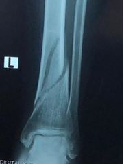 Figure 1: Distal tibia extraarticular fracture
With new treatment approaches, most distal tibia fractures eventually unite. But nonunion has significant
morbidity [8]. It not only affects the limb but has profound impact on person’s mental health and quality of
life [9]. Soft tissue maladaptation and contracture resulting from nonunion at times compromise successful
restoration of limb length or adjacent articular mobility [10]. Patients with a nonunion are 97 times more
likely to need a reoperation [11]. The impact of tibial fracture nonunion on physical health was comparable
with the reported impact of end-stage hip arthrosis and worse than that of congestive heart failure [9]. These
patients also have significant lost productivity resulting in indirect costs [12].
The main target of this review is to identify the factors responsible for the complication (delayed union or
nonunion) of distal tibial fracture in our set up. We have seen increased use of distal locking plates in these
fractures and at the same time we observe many cases of disturbed healing of fractures due to inappropriate
use of fixation device. This review paper clearly brings out the salient points for discussion and understanding.
Methods
A thorough literature search was made in pubmed using key words: extraarticular tibia fractures, distal
tibia fractures, nonunion of distal tibia. Out of 466 articles, case reports, case series and articles published
before 2010 were excluded. Original research articles and review articles published between 2010 January to
2018 December were considered for review. Authors independently reviewed all 241 article abstracts. After
abstract were studied, 51 articles were considered for full text review. This review paper is based on author’s
understanding of relevant literature related to extraarticular fractures of distal tibia.
Conservative Treatment
Conservative treatment of undisplaced distal tibia fractures with cast has showed acceptable functional
outcome in various studies. (Fig. 2) Pandey et al reported a series of cast treatment of nonarticular distal tibia
fractures where all fractures united within 6 months. Fracture displacement in plaster cast and malunion was
observed in few cases, but functional outcome was within acceptable limit [13].
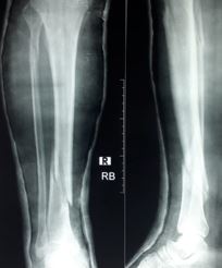 Figure 2: Recurvatum deformity in conservatively treated distal tibia fracture
Sarmiento et al reported that the angular deformity and shortening in distal tibia fractures managed with
functional brace was comparable with those reported by other investigators using intramedullary nails and
functional outcome was excellent [14,15]. Later in 2008, Sarmiento recommended against functional bracing
to fractures that initially have greater than 12mm shortening and angular deformity greater than 8º [16].
External Fixation
Traditionally external fixation has been reserved for trauma with severe skin injury, as a temporary solution
in a two-staged protocol [17] (Fig. 3). Definitive primary external fixation of distal tibia fractures may result
in insufficient reduction, malunion, and pin tract infection.6 Circular external fixator might be preferred due
to its easy application, fewer complications, early mobilisation and shorter treatment time. Illizarov circular
external fixator provides immediate weight bearing as tolerated, irrespective of radiological or clinical healing
with no infection, deformity or non-union [18].
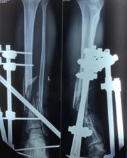 Figure 3: External fixation of open distal tibia fractures
Extracutaneous Locking Compression Plate
External fixation using a precontoured distal femoral locking plate is easy to perform, less invasive, and
the low profile plate can be concealed under stockings [19]. Distal tibial fracture is an ideal indication
for extracutaneous locking plate. The external plating is characterized by fewer soft tissue impingement,
improved cosmesis, and convenient for removal. For optimum stability, the plate-bone distance should be
less than 30mm [20].
Intramedullary Nailing
Intramedullary nailing is the most popular method of treating extraarticular distal tibia fractures. It allows
atraumatic closed stabilization, preserves vascularity of fracture site and integrity of soft tissue envelope and
has fewer wound problems (Fig. 4). Because of the hourglass shape of the medullary canal, it is wider at
fracture site than at the isthmus. It prevents intimate contact between the nail and endosteum. This results in
the play of the nail along the interlocking screws. Torsional and angular stability of fixation is questionable
and there is risk of malunion or nonunion [21-23].
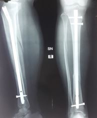 Figure 4: Intramedullary nailing of distal tibia fractures
Intramedullary nailing produces higher rates of malunion in the distal fractures [24]. Distal tibial
malalignment may be more poorly tolerated than proximal malalignment. The incidence of delayed union
or nonunion and implant failure is higher with unreamed nailing, which is attributed to smaller sized nails
[25-27].
Various techniques have been recommended to improve stability of nailing the metaphyseal fractures
including blocking screws (poller screw), temporary unicortical plating, percutaneous reduction clamps, and
fibular plating [28]. Use of blocking (poller) screws in combination with an intramedullary nail can improve
and maintain reduction and fixation of distal tibia fractures at the metaphysio-diaphyseal junction [29].
Adjunctive fibular stabilization in unstable distal tibia fibula fractures helps to prevent malalignment of tibia
nailing [30].
Newer Intramedullary Nails
Expert tibial nail (ETN) has multidirectional locking options. So fibula plating is not necessary with ETN.
This helps to avoid soft tissue problems and delayed healing attributed to fibula fixation [31,32]. Retrograde
tibial nail has produced comparative results in biomechanical comparison with expert tibial nail but is still
in experimental stage [33].
Locking Plate Osteosynthesis
Locking plate osteosynthesis can be used either as a bridge plate, resulting in relative stability or as a
neutralisation plate, in combination with lag screw providing absolute stability [34,35]. Extraarticular distal
tibia fractures fixed with minimally invasive percutaneous plate osteosynthesis (MIPPO) technique using
locking plate have advantages of shorter operation time, less wound complications and less malunion than
with intramedullary nailng. Locking plates have shown union in all cases and very low complications even
in the intraarticular fractures of distal tibia [36,37] (Fig-5).
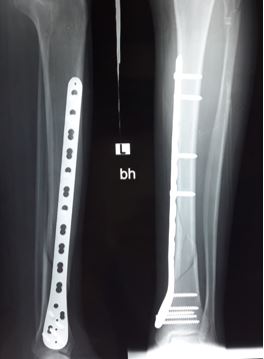 Figure 5: Minimally invasive fixation of distal tibia fracture using locking compression plate
MIPPO respects the soft tissue envelope along with maintaining the biological environment needed for
proper osseous healing [38,39]. Additional use of interfragmentary screws helps to achieve better reduction
and faster fracture healing in simple distal tibia fractures, with fewer complications [34,40]. However, for
complex and comminuted fractures, relative stability with bridge plating by MIPPO is the preferred method
for correcting bone alignment and protecting soft tissue, leading to functional recovery [40-42]. Malleolar
skin irritation is common problem because of prominent hardware.
Though the fracture union rates are similar in locked and nonlocked plating, higher incidence of malalignment
and reoperation are seen with nonlocked plating [43,44].
Intramedullary Nailing or Locking Plate?
However, for metaphyseal fractures or those at the metaphyseal-diaphyseal junction, choice of fixation device
and technique is controversial. Prevalence of valgus malunion was higher in the IMN group and recurvatum
was more prevalent in the MIPO group [45]. Many authors have found no or minor differences in outcome
with nailing or plating techniques [46-51]. Complications in both the groups appeared to be related to
high-energy mechanisms and open fractures [52].
Intramedullary Nail combined with Plate
There are certain scenarios like very distal fracture preventing the required fixation stability and/or a large
bony defect that extends into the metaphysis causing the nail to take a significant more amount of load that
might lead to failure; in these specific scenarios an intramedullary nail plate combination can be extremely
helpful in achieving one’s desired goals in restoring appropriate length, alignment, and rotation [53].
Newer Methods
Various new methods applicable to distal tibial fractures have been studied, including dynamic locking
screws [54], retgrograde tibial nail [55], etc. But their rate of nonunion is yet to be tested in large population.
Once nonunion has been established, low intensity pulsed ultrasound, dynamization [56], reamed exchange
nailing [57,58], expandable intramedullary nail with autologous bone graft [59], and posterolateral plating
with or without bone grafting [60-62] have shown good outcomes in appropriately selected patients,
which is beyond the scope of this review. Whatever treatment planned for nonuninon, should be done after
identification and appropriate management of all modifiable risk factors (e.g. smoking, use of NSAIDs)
[57].
Discussion
It is widely accepted principle for fixation of extraarticular fractures that flexible fixation with moderate
axial movement provides an effective stimulus for periosteal callus formation and thereby accelerates healing
[5]. Whilst axial compression is regarded as stimulatory, the role of interfragmentary shear is controversial
[63]. In a series of cast treatment by Pandey et al, all fractures untied within 6 months. E. Hasenboehler et
al, found prolonged duration required for healing in simple fractures with MIPPO [38]. In another study conducted by Dinko Vidovic, there was delayed union in two patients (9.5%), both of whom had simple
fracture patterns (43 A1) treated with MIPPO [64]. The nail and plate together resulted in better result
whereas the plate and nail singly had no differences with the outcome.
Tissue differentiation at fracture site depends on strain, which in turn depends on the distance between the
moving fracture surfaces [65]. In multifragmentary comminuted fragments, the movement is distributed
between many fragments, so the movement is enough to stimulate callus formation and secondary bone
healing.5 In simple fractures, there are only two moving surfaces. It effects on the formation of cartilage or
bone tissue. Cartilage is formed with less stability and with more stability bone tissue is formed. So it should
be fixed with absolute stability of surgical technique, otherwise it would create delayed union or nonunion
[66] (Fig. 6).
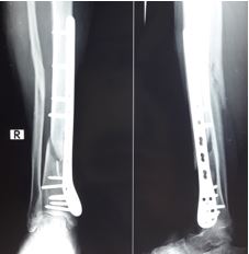 Figure 6: Nonunion of distal tibia following bridge plating of two part fracture
In clinical practice, the majority of nonunions are due to mechanical problems with instability, resulting in
too much strain at the fracture site. The bone maintains its biological potential to heal, but fails to function
due to the mechanical conditions. The majority of nonunions will heal if the correct mechanical environment
is produced by surgery, without the need for biological adjuncts such as autologous bone graft [67]. It is
important however, to preserve soft tissue envelope.
Conclusion
The treatment of distal tibia fractures is controversial. We prefer absolute stability in two part fractures to
eliminate interfragmentary shear force; and relative stability in multifragmentary comminuted fractures.
Choice of fixation device depends on availability of implant and expertise of surgeon.
Bibliography
- Calori, G. M. & Giannoudis, P. V. (2011). Enhancement of fracture healing with the diamond concept: the role of the biological chamber. Injury, 42(11), 1191-1193.
- Rodriguez-Buitrago, A. F. & Jahangir, A. (2018). Tibia Nonunion. StatPearls. Treasure Island (FL): StatPearls Publishing StatPearls Publishing LLC.
- Mills, L., Tsang, J., Hopper, G., Keenan, G. & Simpson, A. H. (2016). The multifactorial aetiology of fracture nonunion and the importance of searching for latent infection. Bone Joint Res., 5(10), 512-519.
- Augat, P., Simon, U., Liedert, A. & Claes, L. (2005). Mechanics and mechano-biology of fracture healing in normal and osteoporotic bone. Osteoporos Int., 16(Suppl 2), S36-43.
- Claes, L. E., Heigele, C. A., Neidlinger-Wilke, C., Kaspar, D., Seidl, W., Margevicius, K. J., et al. (1998). Effects of mechanical factors on the fracture healing process. Clin Orthop Relat Res., (355 Suppl), S132-47.
- Zelle, B. A., Bhandari, M., Espiritu, M., Koval, K. J., Zlowodzki, M. & Group obotE-BOTW (2006). Treatment of Distal Tibia Fractures Without Articular Involvement: A Systematic Review of 1125 Fractures. Journal of Orthopaedic Trauma, 20(1), 76-79.
- Achten, J., Parsons, N. R., McGuinness, K. R., Petrou, S., Lamb, S. E., Costa, M. L. (2015). UK Fixation of Distal Tibia Fractures (UK FixDT): protocol for a randomised controlled trial of 'locking' plate fixation versus intramedullary nail fixation in the treatment of adult patients with a displaced fracture of the distal tibia. BMJ Open., 5(9), e009162.
- Elniel, A. R. & Giannoudis, P. V. (2018). Open fractures of the lower extremity: Current management and clinical outcomes. EFORT Open Rev., 3(5), 316-325.
- Brinker, M. R., Hanus, B. D., Sen, M. & O'Connor, D. P. (2013). The devastating effects of tibial nonunion on health-related quality of life. J Bone Joint Surg Am., 95(24), 2170-2176.
- Ring, D., Barrick, W. T. & Jupiter, J. B. (1997). Recalcitrant nonunion. Clin Orthop Relat Res., (340), 181-189.
- Johnson, K. D. (1987). Management of malunion and nonunion of the tibia. Orthop Clin North Am., 18(1), 157-171.
- Tay, W. H., de Steiger, R., Richardson, M., Gruen, R. & Balogh, Z. J. (2014). Health outcomes of delayed union and nonunion of femoral and tibial shaft fractures. Injury, 45(10), 1653-1658.
- Pandey, B. K., Manandhar, R. R., Sharma, S., Pradhan, R. L., Lakhey, S. & Rijal, K. P. (2009). Conservative treatment of nonarticular fractures of distal third tibia. JNMA J Nepal Med Assoc., 48(176), 292-295.
- Sarmiento, A. & Latta, L. L. (2004). 450 closed fractures of the distal third of the tibia treated with a functional brace. Clin Orthop Relat Res., (428), 261-271.
- Sarmiento, A. & Latta, L. L. (1999). Functional fracture bracing. J Am Acad Orthop Surg., 7(1), 66-75.
- Sarmiento, A. & Latta, L. L. (2008). Fractures of the middle third of the tibia treated with a functional brace. Clin Orthop Relat Res., 466(12), 3108-3115.
- Joveniaux, P., Ohl, X., Harisboure, A., Berrichi, A., Labatut, L., Simon, P., et al. (2010). Distal tibia fractures: management and complications of 101 cases. Int Orthop., 34(4), 583-588.
- Fadel, M., Ahmed, M. A., Al-Dars, A. M., Maabed, M. A., Shawki, H. (2015). Ilizarov external fixation versus plate osteosynthesis in the management of extra-articular fractures of the distal tibia. Int Orthop., 39(3), 513-519.
- Zhang, J., Ebraheim, N. A., Li, M., He, X., Liu, J., Zhu, L., et al. (2015). External Fixation Using a Locking Plate: A Reliable Way in Treating Distal Tibial Fractures. J Orthop Trauma., 29(11), e454-8.
- Zhang, J., Ebraheim, N., Li, M., He, X., Schwind, J., Liu, J., et al. (2015). External fixation using locking plate in distal tibial fracture: a finite element analysis. Eur J Orthop Surg Traumatol., 25(6), 1099-1104.
- Bedi, A., Le, T. T. & Karunakar, M. A. (2006). Surgical treatment of nonarticular distal tibia fractures. J Am Acad Orthop Surg., 14(7), 406-416.
- Drosos, G., Karnezis, I. A., Bishay, M. & Miles, A. W. (2001). Initial rotational stability of distal tibial fractures nailed without proximal locking: the importance of fracture type and degree of cortical contact. Injury, 32(2), 137-143.
- Neumann, M. V., Strohm, P. C., Reising, K., Zwingmann, J., Hammer, T. O. & Suedkamp, N. P. (2016). Complications after surgical management of distal lower leg fractures. Scand J Trauma Resusc Emerg Med., 24(1), 146.
- Ehlinger, M., Adam, P., Gabrion, A., Jeunet, L., Dujardin, F. & Asencio, G. (2010). Distal quarter leg fractures fixation: The intramedullary nailing alone option. Orthop Traumatol Surg Res., 96(6), 674-682.
- Duan, X., Al-Qwbani, M., Zeng, Y., Zhang, W. & Xiang, Z. (2012). Intramedullary nailing for tibial shaft fractures in adults. Cochrane Database Syst Rev., 1, Cd008241.
- Blachut, P. A., O'Brien, P. J., Meek, R. N. & Broekhuyse, H. M. (1997). Interlocking intramedullary nailing with and without reaming for the treatment of closed fractures of the tibial shaft. A prospective, randomized study. J Bone Joint Surg Am., 79(5), 640-646.
- Weninger, P., Schueller, M., Jamek, M., Stanzl-Tschegg, S., Redl, H. & Tschegg, E. K. (2009). Factors influencing interlocking screw failure in unreamed small diameter nails--a biomechanical study using a distal tibia fracture model. Clin Biomech (Bristol, Avon)., 24(4), 379-384.
- Moongilpatti Sengodan, M., Vaidyanathan, S., Karunanandaganapathy, S., Subbiah Subramanian, S. & Rajamani, S. G. (2014). Distal tibial metaphyseal fractures: does blocking screw extend the indication of intramedullary nailing? ISRN Orthop., 2014(542623), 1-7.
- Shah, R. K. & Shah, S. B. (2015). Treatment of Diaphysio-Metaphyseal Fracture of Tibia by Intramedullary Nail in Combination with Poller Screw. Journal of Bone Research and Reports, 1(1).
- Egol, K. A., Weisz, R., Hiebert, R., Tejwani, N. C., Koval, K. J. & Sanders, R. W. (2006). Does fibular plating improve alignment after intramedullary nailing of distal metaphyseal tibia fractures? J Orthop Trauma., 20(2), 94-103.
- El Attal, R., Hansen, M., Rosenberger, R., Smekal, V., Rommens, P. M. & Blauth, M. (2011). [Intramedullary nailing of the distal tibia illustrated with the Expert(TM) tibia nail]. Oper Orthop Traumatol., 23(5), 397-410.
- Wang, Z., Cheng, Y., Xin, D., Liu, T., Qu, W., Wang, D., et al. (2017). Expert Tibial Nails for Treating Distal Tibial Fractures with Soft Tissue Damage: A Patient Series. J Foot Ankle Surg., 56(6), 1232-1235.
- Kuhn, S., Appelmann, P., Pairon, P., Mehler, D., Hartmann, F. & Rommens, P. M. (2014). A new angle stable nailing concept for the treatment of distal tibia fractures. Int Orthop., 38(6), 1255-1260.
- Wenger, R., Oehme, F., Winkler, J., Perren, S. M., Babst, R. & Beeres, F. J. P. (2017). Absolute or relative stability in minimal invasive plate osteosynthesis of simple distal meta or diaphyseal tibia fractures? Injury, 48(6), 1217-1223.
- Egol, K. A., Kubiak, E. N., Fulkerson, E., Kummer, F. J. & Koval, K. J. (2004). Biomechanics of locked plates and screws. Journal of orthopaedic trauma, 18(8), 488-493.
- Shukla, R., Jain, N., Jain, R. K., Patidar, S. & Kiyawat, V. (2018). Minimally Invasive Plate Osteosynthesis Using Locking Plates for AO 43-Type Fractures: Lessons Learnt From a Prospective Study. Foot Ankle Spec., 11(3), 236-241.
- Vidovic, D., Matejcic, A., Ivica, M., Jurisic, D., Elabjer, E. & Bakota, B. (2015). Minimally-invasive plate osteosynthesis in distal tibial fractures: Results and complications. Injury, 46(Suppl 6), S96-9.
- Hasenboehler, E., Rikli, D. & Babst, R. (2007). Locking compression plate with minimally invasive plate osteosynthesis in diaphyseal and distal tibial fracture: a retrospective study of 32 patients. Injury, 38(3), 365-370.
- Lai, T. C. & Fleming, J. J. (2018). Minimally Invasive Plate Osteosynthesis for Distal Tibia Fractures. Clin Podiatr Med Surg., 35(2), 223-232.
- Li, Q., Zeng, B. F., Luo, C. F., Song, S., Zhang, C. Q. & Kong, W. Q. (2014). Limited open reduction is better for simple- distal tibial shaft fractures than minimally invasive plate osteosynthesis. Genet Mol Res., 13(3), 5361-5368.
- Qi, H., Li, W., Zhao, Y., Zhang, Y., Liu, Z. & Jia, J. (2013). [Comparison study on two operations for treatment of extra-articular distal tibial fracture]. Zhongguo Xiu Fu Chong Jian Wai Ke Za Zhi., 27(11), 1286-1290.
- Zhang, Q. X., Gao, F. Q., Sun, W., Wang, Y. T., Yang, Y. R. & Li, Z. (2015). [Minimally invasive percutaneous plate osteosynthesis versus open reduction and internal fixation for distal tibial fractures in adults: a meta-analysis]. Zhongguo Gu Shang, 28(8), 757-762.
- Hahn, D., Bradbury, N., Hartley, R. & Radford, P. J. (1996). Intramedullary nail breakage in distal fractures of the tibia. Injury, 27(5), 323-327.
- Ahmad, M. A., Sivaraman, A., Zia, A., Rai, A., Patel, A. D. (2012). Percutaneous locking plates for fractures of the distal tibia: our experience and a review of the literature. J Trauma Acute Care Surg., 72(2), E81-7.
- Beytemur, O., Baris, A., Albay, C., Yuksel, S., Caglar, S. & Alagoz, E. (2017). Comparison of intramedullary nailing and minimal invasive plate osteosynthesis in the treatment of simple intra-articular fractures of the distal tibia (AO-OTA type 43 C1-C2). Acta Orthop Traumatol Turc., 51(1), 12-16.
- Tejwani, N., Polonet, D. & Wolinsky, P. R. (2014). Controversies in the intramedullary nailing of proximal and distal tibia fractures. J Am Acad Orthop Surg., 22(10), 665-673.
- Tejwani, N. C., Polonet, D. & Wolinsky, P. R. (2015). Controversies in the intramedullary nailing of proximal and distal tibia fractures. Instr Course Lect., 64, 175-183.
- Fang, J. H., Wu, Y. S., Guo, X. S. & Sun, L. J. (2016). Comparison of 3 Minimally Invasive Methods for Distal Tibia Fractures. Orthopedics, 39(4), e627-33.
- Kuo, L. T., Chi, C. C. & Chuang, C. H. (2015). Surgical interventions for treating distal tibial metaphyseal fractures in adults. Cochrane Database Syst Rev., (3), Cd010261.
- Newman, S. D. S., Mauffrey, C. P. C. & Krikler, S. (2011). Distal metadiaphyseal tibial fractures. Injury, 42(10), 975-984.
- Iqbal, H. J. & Pidikiti, P. (2013). Treatment of distal tibia metaphyseal fractures; plating versus intramedullary nailing: a systematic review of recent evidence. Foot Ankle Surg., 19(3), 143-147.
- Barcak, E. & Collinge, C. A. (2016). Metaphyseal Distal Tibia Fractures: A Cohort, Single-Surgeon Study Comparing Outcomes of Patients Treated with Minimally Invasive Plating Versus Intramedullary Nailing. J Orthop Trauma., 30(5), e169-74.
- Yoon, R. S. & Liporace, F. A. (2016). Intramedullary Nail and Plate Combination Fixation for Complex Distal Tibia Fractures: When and How? J Orthop Trauma., 30(Suppl 4), S17-S21.
- Acklin, Y. P., Stockle, U. & Sommer, C. (2016). Clinical and radiologic outcomes associated with the use of dynamic locking screws (DLS) in distal tibia fractures. Eur J Trauma Emerg Surg., 42(3), 351-356.
- Kuhn, S., Appelmann, P., Pairon, P., Mehler, D. & Rommens, P. M. (2014). The Retrograde Tibial Nail: Presentation and biomechanical evaluation of a new concept in the treatment of distal tibia fractures. Injury, 45(Suppl 1), S81-S86.
- Vaughn, J., Gotha, H., Cohen, E., Fantry, A. J., Feller, R. J., Van Meter, J., et al. (2016). Nail Dynamization for Delayed Union and Nonunion in Femur and Tibia Fractures. Orthopedics, 39(6), e1117-e23.
- Tsang, S. T., Mills, L. A., Frantzias, J., Baren, J. P., Keating, J. F. & Simpson, A. H. (2016). Exchange nailing for nonunion of diaphyseal fractures of the tibia: our results and an analysis of the risk factors for failure. Bone Joint J., 98-B(4), 534-541.
- Hierholzer, C., Friederichs, J., Glowalla, C., Woltmann, A., Buhren, V. & von Ruden, C. (2017). Reamed intramedullary exchange nailing in the operative treatment of aseptic tibial shaft nonunion. Int Orthop., 41(8), 1647-1653.
- Niu, Y., Bai, Y., Xu, S., Liu, X., Wang, P., Wu, D., et al. (2011). Treatment of lower extremity long bone nonunion with expandable intramedullary nailing and autologous bone grafting. Arch Orthop Trauma Surg., 131(7), 885-891.
- Foster, M. J., O'Toole, R. V. & Manson, T. T. (2017). Treatment of tibial nonunion with posterolateral bone grafting. Injury, 48(10), 2242-2247.
- Konda, S., Saleh, H., Fisher, N. & Egol, K. A. (2017). Posterolateral Bone Grafting for Distal Tibia Nonunion. J Orthop Trauma., 31(Suppl 3), S16.
- Lin, C., Lin, L., Vinesh, L., Shao, X., Lu, X. & Hong, J. (2017). Distal tibial nonunion using a contralateral anterior L-shaped locking compression plate through a posterior-lateral approach: A retrospective case series. Injury, 48(6), 1224-1228.
- Epari, D. R., Taylor, W. R., Heller, M. O. & Duda, G. N. (2006). Mechanical conditions in the initial phase of bone healing. Clin Biomech (Bristol, Avon)., 21(6), 646-655.
- Richard, R. D., Kubiak, E. & Horwitz, D. S. (2014). Techniques for the surgical treatment of distal tibia fractures. Orthop Clin North Am., 45(3), 295-312.
- Perren, S. M. (2010). [Optimizing the degree of fixation stability based on the strain theory]. Orthopade, 39(2), 132-138.
- Shen, J., Xu, J., Tang, M. J., Luo, C. F. & Zhang, C. Q. (2016). Extra-articular distal tibia facture (AO-43A): A retrospective study comparing modified MIPPO with IMN. Injury, 47(10), 2352-2359.
- Elliott, D. S., Newman, K. J., Forward, D. P., Hahn, D. M., Ollivere, B., Kojima, K., et al. (2016). A unified theory of bone healing and nonunion: BHN theory. Bone Joint J., 98-B(7), 884-891.

