Biography
Interests
Carlos Vilchez, A. S.1, Nina Abreu, M. P.1, Pablo Coimbra, P. A.1, João Rodrigues, P. C.1, Caio Malveira1, Fernando Carvalho, M.1, Laura Gomes, V. M.2 & Antonio Junior, G. L.1*
1Radiology Unit, Hospital Antonio Prudente, Fortaleza, CE, Brazil
2UniRV, Goianésia, GO, Brazil
*Correspondence to: Dr. Antonio Junior, G. L., Radiology Unit, Hospital Antonio Prudente, Fortaleza, CE, Brazil.
Copyright © 2021 Dr. Antonio Junior, G. L., et al. This is an open access article distributed under the Creative Commons Attribution License, which permits unrestricted use, distribution, and reproduction in any medium, provided the original work is properly cited.
Abstract
Bilateral cavernous carotid fistulas are rare, it is an abnormal vascular shunt from the carotid artery
to the cavernous sinus.
We report a case of a 55-years-old woman, who developed Barrow’s type D carotid-cavernous
fistula.
Magnetic resonance imaging is of great importance for the diagnosis.
Background
Carotid-cavernous fistulas are characterized by abnormal connections between the carotid circulation and
the cavernous sinus. The clinical picture is very wide, ranging from pulsatile exophthalmos to intracranial
bleeding. Fistulas can be classified as direct or indirect, both being different conditions with different
etiologies. The study aims to show a case where it was possible to suspect the carotid-cavernous fistula with
indirect signs on magnetic resonance, with subsequent confirmation by angiographic study.
Case Report
A female patient, 55 years old, about 30 years ago, started strabismus associated with diplopia and bilateral
exophthalmos. Submitted to a magnetic resonance imaging of the orbits and skull that showed indirect
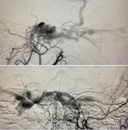
signs of bilateral carotid-cavernous fistula, then being referred for cerebral angiography, which confirmed
Barrow’s type D carotid-cavernous fistula.
Discussion
Carotid-cavernous fistulas are characterized by abnormal connections between the carotid circulation and
venous drainage, more specifically the cavernous sinus. Among the findings of the clinical picture, pulsatile
proptosis, progressive visual loss, subarachnoid hemorrhage, intracerebral hemorrhage, and involvement
of several pairs of cranial nerves (III, IV, V, and VI) are observed. The radiological findings on magnetic
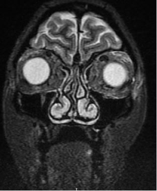
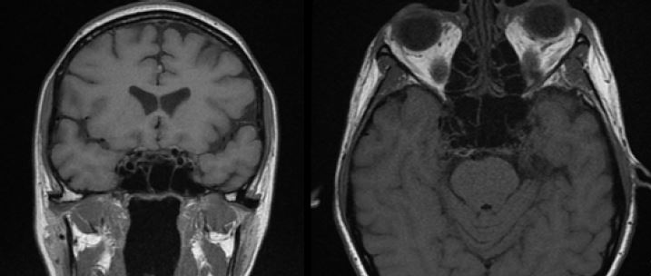
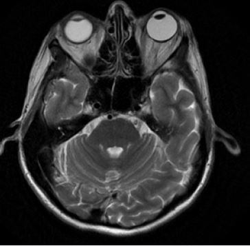
resonance imaging that made it possible to suspect the condition was engorgement and enlargement of the left
cavernous sinus with prominent flow-void at the site, associated with dilation of the upper ophthalmic vein,
proptosis of the bilateral eyeball, and edema of the left periorbital fat. Other radiological findings reported
in the literature are anomalous contrast enhancement and asymmetric enhancement of the cavernous sinus
and dehiscent carotid artery. There are several classifications for the pathology, however the most important
is related to flow intensity, vascular anatomy, and Barrow’s angiographic classification. Regarding vascular
anatomy, they are divided into direct and indirect. In the first, communication between the internal carotid
artery and the cavernous sinus occurs directly, while in the indirect fistula, there is communication through
branches of the carotid circulation. There is also the Barrow angiographic classification characterized in:
A: a connection between the intracavernous internal carotid artery and the cavernous sinus; B dural shunt
between intracavernous branches of the internal carotid and cavernous sinus; C: dural shunts between
meningeal branches of the external carotid artery and cavernous sinus; D: mixed with components B and C.
The therapeutic approach is varied and there may be conservative management [1-4].
Conclusions
Although the angiographic study is essential in confirming the diagnosis and therapeutic planning, analysis
by magnetic resonance imaging is extremely important, as several radiological findings allow the clinical
suspicion of carotid-cavernous fistula.
Acknowledgements
None.
Authors’ Contributions
CV and AJ designed the study, acquired and interpreted the data, and have to be personally accountable for
the accuracy and integrity of the entire work. NA, PC, JR, CM and JM provided clinical care to the patient,
performed literature searches, interpreted the data, and drafted the manuscript. LG collected, analyzed,
interpreted the data, study design and conception. All authors reviewed and revised the manuscript and
approved the final manuscript.
Funding
None.
Availability of Data and Materials
The datasets used and/or analysed during the current study are available from the corresponding author on
reasonable request.
Ethics Approval and Consent to Participate
The patient was consented for participation in the study.
Consent for Publication
The patient is aware of this case report submission and has provided written consent for this publication.
Competing Interests
The authors report no competing interests
Bibliography


Hi!
We're here to answer your questions!
Send us a message via Whatsapp, and we'll reply the moment we're available!