Biography
Interests
Mahendra Pal1* & Pratibha Dave2
1Narayan Consultancy on Veterinary Public Health and Microbiology, Aangan, Jagnath Ganesh Dairy Road,
Gujarat, India
2Welfare Hospital and Research Centre, Bharauch, Gujarat, India
*Correspondence to: Dr. Mahendra Pal, Narayan Consultancy on Veterinary Public Health and Microbiology, Aangan, Jagnath Ganesh Dairy Road, Gujarat, India.
Copyright © 2019 Dr. Mahendra Pal and Dr. Pratibha Dave. This is an open access article distributed under the Creative Commons Attribution License, which permits unrestricted use, distribution, and reproduction in any medium, provided the original work is properly cited.
Abstract
Mycetoma is a neglected, chronic, progressive, granulomatous disease of the skin and subcutaneous tissues, which sometimes involves the muscle, bone, and neighboring tissues. Mycetoma continues to pose a huge public health threat in many tropical and sub-tropical countries of the world. It is caused by bacteria (actinomycetoma) as well as fungi (eumycetoma), which occur in the saprobic environment. The soil serves as the reservoir of infection and transmission occurs through skin inoculation of the pathogen following trauma. The prevalence of etiological agents of mycetoma varies from region to region. In Brazil and Mexico, Nocardia brasiliensis is the main etiologic agent of actinomycetoma. Madurella mycetomatis is most common cause of eumycetoma in Africa. The bacteria accounts for about 60% and fungi for 40% of mycetoma in the world. Disease is more prevalent in developing countries as compared to the developed nations. Mycetoma is observed more frequently in men than women. The maximum cases are recorded in young adults in the age group of 20 to 40 years. The incidence of mycetoma is higher in rural areas than in urban settings. In endemic areas, the incidence of mycetoma is more among the persons who walk barefoot. It is considered as an occupational disease of farmers, builders, gardeners, field employees, carpenters, herdsmen, and land workers who are at greater risk of traumatic injury providing an opportunity for the entry of the causative agents. In the absence of reliable immunological technique, microbiological, histopathological, and molecular tests are employed to confirm the diagnosis of disease. Mycetoma should be differentiated from actinomycosis, botryomycosis, dematophytosis, elephantiasis, leprosy, sporotrichosis, tuberculosis, and benign tumors. Antibacterial antibiotics and antifungal drugs are used for the management of actinomycetoma and eumycetoma, respectively. Recurrence is commonly observed in eumycetoma. Currently, no vaccine is available; and therefore, people who work with soil related activities are advised to wear protective clothing and seek immediate medical help after the skin injury.
Introduction
The neglected diseases are caused by a variety of pathogens, which include viruses, bacteria, fungi, protozoa,
helminthes, and ectoparasites [1]. Many neglected diseases, such as anthrax, bovine tuberculosis, brucellosis,
Chagas disease, chickungunya, chromobalstomycosis, echinococcosis, dengue, dracunculiasis, lymphatic
filariasis, leishmaniasis, leprosy, leptospirosis, malaria, mycetoma, onchocercariasis, plague, rabies, scabies,
schistosomiasis, sleeping sickness, taeniasis/cysticercosis, trachoma, and yaws, are reported in poor people
living in developing nations of the world [2,3]. According to the World Health Organization (WHO),
about one billion people in 198 countries are affected with neglected tropical diseases [2]. Mycetoma is
recently included in the list of tropical neglected diseases by WHO in 2017. The word "mycetoma" means
"fungus tumor" that is caused by aerobic actinomycetes and saprobic fungi, which occur in the environment.
The name “Madura foot” originates from the place “Madurai” of South India where it was first noticed in
1842 by Gill. The role of fungi in the etiology of mycetoma was recognized by Carter in 1860. Mycetoma
(Madura foot, maduromycosis) is a localized, suppurative, granulomatous, infectious, chronic debilitating
disease, which is described from many countries of the world, such as Argentina, Brazil, Chad, Columbia,
Djibouti, Egypt, Ethiopia, England, France, Germany, Japan, India, Iran, Lebanon, Mexico, Philippines,
Netherlands, Niger, Nigeria, Saudi Arabia, Senegal, Somalia, Sri Lanka, Sudan, Thailand, Turkey, USA,
Yemen, Venezuela, and others [1,4-9]. Mycetoma is public health issue as it has socioeconomic impact
on patient. Mycetoma is not a reportable disease; and therefore, the true incidence and prevalence is not
known. In African continent, Sudan is considered as the homeland of mycetoma, as annual number of cases
diagnosed in hospitals vary between 300 to 400 [2]. It is considered to be a disease of barefooted people, and
the lesions are usually observed on the foot (70%). Affected foot shows gross swelling, multiple nodules, and
sinuses that yield granules [5].
Madurella mycetomatis is the most common cause of eumyetoma in the world. Clinical diagnosis must be confirmed by laboratory tests, which include microbiological, histopathological, and molecular techniques. Treatment of mycetoma depends on the etiology of the causative agent. Antifungal drugs, such as ketoconazole, itraconazole, and voriconazole are tried for eumycetoma where as actinomycetoma is treated with courses of antibacterial antibiotics like co-trimoxazole, minocycline, amikacin, and refampicin [5,10]. The therapeutic agents used in eumycetoma can show resistance, and therefore, may require surgical interventions including amputation. The present communication focuses on the etiology, epidemiology, diagnosis, and management of mycetoma.
Etiology
Mycetoma is a chronic mutilating disease of the skin and the underlying tissues caused by bacteria
(Actinomadura madurae, Actinomadura pelletieri, Nocardia asteroides, N. brasiliensis, N. harenae, N.
otitidiscaviarum, and N. takedensis, Streptomyces somaliensis), and fungi (Acremonium falciforme, A. kiliense, A.
recifei, A. strictum, Aspergillus nidulans, A. terreus, Cladophialophora bantiana, Curvularia geniculata, C. lunata,
Cylindrocarpon nescens, C. destructans, Exophiala jeanselmei, Fusarium moniliformis, F. soalni, Leptosphaeria
senegalensis, L. tompkinsii, Madurella grisea, M. mycetomatis, Noetestudina rosatii, Phaeoacremonium krajdenii,
Phialophora verrucosa, Pyrenochaeta mackinnonii, P. romeroi, Scedosporium apiospermum (the anamorph or
asexual form of Pseudallescheria boydii), and Pyrenochaeta romeroi) [4-8,10]. These organisms are widely
prevalent in saprobic environment, and are recovered from diverse types of natural substrates including the
soil, excreta, and plants [5]. The role of other species of bacteria and fungi in the etiology of mycetoma needs
to be further investigated.
Transmission
The source of infection is exogenous, as the causative agents of mycetoma (bacteria and fungi) are found in
the soil and other environmental materials. The organisms enter the subcutaneous tissue usually through
minor trauma, lacerated wound or penetrating injury due to actinomycetes and fungal contaminated objects,
such as the thorn, wooden splinter, metal piece, wire, brick etc., [5]. Hitherto, it is not known why some
individuals develop mycetoma and others do not. The role of invertebrate vector or animal in the spread of
infection is not yet elucidated.
Clinical Spectrum
The incubation period of mycetoma is very long that may vary from 3 months to 9 years. Disease presents
with localized tissue swelling, abscess formation, development of sinus tracts, and discharge of grains [2].
Clinical appearance of lesions on the affected body parts of patients appears same both in actinomycetoma
(Fig.1) and eumycetoma (Fig.2). Mycetoma commonly involves the extremities, but other part of the body
can be affected. Mycetoma often begins as a small, painless, subcutaneous nodule at the site of injury, usually
the foot. Gradually, multiple nodules develop, ulcerate, and drain through sinus tracts. The discharge may
be serosanguineous, seropurulent or purulent, and often contains the characteristic granules [5]. When the
disease progresses further, the surrounding tissue swells and gets deformed by fibrous tissue reaction and
multiple sinus formation. Pain is a main complaint when bone involvement or secondary bacterial infection
occurs. Mycetoma of the thorax, head, neck or thorax can lead to death because it can spread into adjacent
vital organs [2].
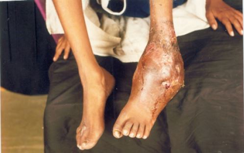
Source: Pal (2007)
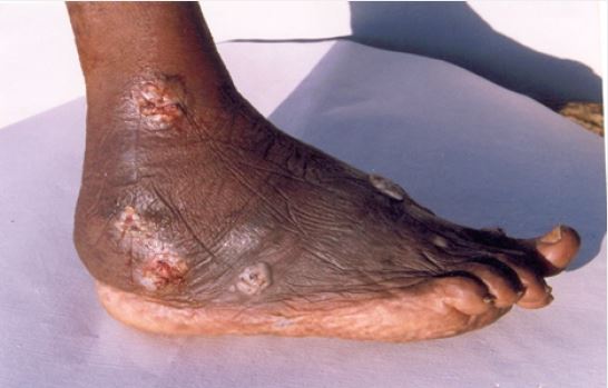
Source: Pal (2007)
Mycetoma typically remains localized, extending slowly, and invading the subcutaneous, fat, ligaments, muscles, and bones but sparing the tendons until very late. The extent and degree of bone involvement varies with the species involved of the etiologic agent. In eumycetoma, the lesion takes the form of single or multiple punched out lytic areas with well defined walls and little sign of bone reaction. In actinomycetoma, both oesteolytic and oesteosclerotic changes are present at the same time. Destruction of the ligaments and articular surfaces results in ankylosis of the joints. Visceral invasion may occur by contiguity when the infection is localized in the head, neck, chest, and buttocks. Extensive fibrosis in the tissue may cause elephantiasis [5].
Epidemiology
Global burden of mycetoma is not known as it is not a notifiable disease. Disease has been known for a long
time in India. It is reported from many developing as well as developed nations of the world [2,6-9,11].
Mycetoma is a public health problem in equatorial regions of Africa, Latin America, and Asia, which are
known as the “mycetoma belt”. Fungal mycetoma (eumycetoma) is the most common type in Africa, while
bacterial mycetoma (actinomycetoma) causes most cases in South and Central America and some Asian
countries. Transmission of infection occurs when the causative organism enters the body through minor
trauma or a penetrating injury, commonly thorn pricks. There is a clear association between mycetoma and
individuals who walk barefooted and are manual workers. Hitherto, there is no evidence of transmission of
infection from human to human or from animal to human.
The disease is sporadic in occurrence; and is not contagious, as there is no record of transmission of the infection between humans or from animals to humans. The foot is the most common site of infection because of its liability to repeated trauma. The infection is rarely seen in children. However, the adult males between 20-40 years are most frequently affected [5]. Mycetoma is generally encountered in rural areas than urban areas due to the nature of work in the field. In endemic regions of Africa and Asia, the incidence of mycetoma is more among the persons who walk barefooted. Certain occupational groups, like farmers, laborers, herdsmen, field workers, and gardeners, etc. who are exposed to the soil and vegetation following skin injuries, are commonly affected [5,10].
Eumycetoma is more commonly observed in Africa, where as actinomycetoma is more frequently encountered in Mexico and Brazil. In South India, actinomycetoma is more prevalent whereas the eumycetoma is more common in North India. The species of the organism causing mycetoma vary from country to country to country. In Sudan, M. mycetomatis has been implicated in more than 70% of cases. Madurella grisea is most frequently observed in the New World. In India, eumycetoma is principally caused by M. mycetomatis. Streptomyces somalensis is frequently found to be implicated with mycetoma in Middle East and Sudan. However, in Mexico and Central America, nocardial mycetoma is most commonly encountered [2].
Diagnosis
Diagnosis is made clinically from observation of the triad of a swollen nodular skin/subcutaneous lesion,
sinus tracts, and grain production. X-rays, computed tomography, and magnetic resonance imaging (MRI)
are useful to detect the extent of the osteolytic activity. Direct microscopical examination of pus reveals the form, colour and size of the grains. Red grains are due to an actinomycete whereas black granules are
observed in fungal mycetoma. The final diagnosis requires the isolation of the etiological agent from the
granules or biopsy materials on cultural media [2,5].
Actinomycetic granules should be repeatedly washed with sterile physiological saline and inoculated on Lowenstein-Jensen medium, blood agar, thioglycollate broth and cooked meat broth. Antibiotic solution containing streptopenicillin or chloramphenico is used to wash the eumycotic granules and then cultured on mycological media, such as Sabouraud medium, Czapek dox agar, Pal sunflower seed medium [12], and APRM (Anubha, Pratibha, Raj, Mahendra) agar [1]. A deep biopsy is an ideal specimen for the culture of the organism, as it is free from external contamination. The organism can be demonstrated in the histopathological section of the biopsy tissue by haemotoxylin and eosin, Kinyoun’s, Brown and Brenn’s, Masson Fontena silver stain, periodic acid Schieff (PAS), and Gomori methanamine silver (GMS) techniques [5]. Molecular tools like polymerase chain reaction (PCR) and loop mediated isothermal amplification (LAMP) are also very useful to diagnose disease. The detailed morphology of the fungal isolates can be easily studied in Narayan stain, which is developed by Pal [13]. It is imperative to differentiate mycetoma from other diseases, such as blastomycosis, botryomycosis, chromoblastomycosis, coccidioidomycosis, nocardiosis, sporotrichosis, and tuberculosis by employing standard microbiological techniques [10]. As Narayan stain is cheaper than other staining solutions, therefore, it should be routinely used in the laboratory of public health and microbiology for morphological studies of fungi, which are implicated in many clinical disorders of humans as well as animals.
Treatment
Mycetoma poses a challenge in treatment. The therapy of mycetoma depends primarily on the causative
agent, site of infection, and extent of the disease. Chemotherapy has proved to be helpful in early cases and
should be continued for several months after clinical cure to prevent relapse. Actinomycetoma is generally
treated with therapeutic agents [2,14]. However, a combination of medical treatment and surgical excision
of affected part is the gold standard for the management of eumycetoma [2,15,16].
In actinomycetoma, dapsone (200-300mg daily for 6-24 months), co-trimoxazole 2 tablets (480mg twice daily for several months), amikacin (500mg intramuscularly daily for 3 months followed by a two weeks rest period and repeated), doxycycline (100mg twice daily for 6-12 months), sulphadiazine (3-8g/day for 6-9 months), minocycline (150mg twice a day for few months), penicillin (500mg orally four times daily for 3 months) have been used [5]. A combination of sulphadoxine and pyrimethamine or streptomycin and refampicin or co-trimaxazole and dapsone or dapsone with amikacin and ampicillin and chloramphenicol or tetracycline has also been tried [2,5,15]. Ciprofloxan has been effective in treating actinomycetma with bone involvement. The patients who showed resistance to amikacin can be treated with netilmicin [2].
Treatment modalities of eumycetomas include amphotericin B (0.5-1.2mg/kg/day), Iitraconazole (200- 400mg/day), ketoconazole (400mg/day), posaconazole (200mg, four times daily), terbinafine (500-1000mg/ day), and voriconazole 400-600mg/day, alone or with any combination(s). Treatment is typically given for months to years. Recently, Dave and Pal (2015) [10] treated a case of mycetoma due to Curvularia lunata in a 45-year-old gardener with itraconazole (200mg daily for two months). The drug was well tolerated and no side effects were noticed during the course of therapy. The eumycetes are generally resistant to antifungal drugs, and may require surgical interventions including amputation. Eumycetoma has a poor prognosis, with only a 40% rate of cure. It is emphasized that the clinical efficacy of fosravuconazole and isavuconazole should be assessed in eumycetomas.
Surgical treatment can be considered either as an adjunct to medical therapy or when all attempts to control the infection have failed. Localized lesions, which can be excised without residual disability, are best treated. Amputation is considered as last measure if the lesion becomes too large, and does not respond to conservative line of treatment and causes severe disability [5].
The prognosis of disease differs according to the etiological agents. It is pertinent to mention that irregular or incomplete course of therapy can result relapse of infection [2]. The postoperative recurrence rate may vary from 25% to 50% [15].
Prevention
Currently, no vaccine is available to immunize the susceptible population in endemic regions of the world
[10]. The person should avoid penetrating wounds by thorns and splinters. Immediate attention should be
paid to the traumatic skin lesions to prevent implantation of infectious organisms from saprobic material.
Certain occupation groups, such as gardeners, agricultural workers, farm laborers, and other persons who
have frequent contact with the soil and vegetations, should wear shoes and other protective clothing [5]. It
is advised that people living in the endemic areas should not walk barefooted [2].
Conclusion
Mycetoma, a neglected infectious disease, is reported from tropical, subtropical and also temperate regions
of the world. Disease is caused by fungi and bacteria, which exist as saprophytes in the environment.
Trauma is the chief predisposing factor for the initiation of infection. The source of infection is exogenous,
and the organisms usually enter the body following injury to the skin. The route of infection is not well
understood. The disease is characterized by tumefaction, draining sinuses, and the presence of grains. The
lesion commonly occurs in the foot. Disease usually affects the poor communities in remote area of many
countries of the world including India. Mycetoma affects people of all ages and is more common in men.
People, who work in agricultural related jobs, are at a greater risk of getting the infection. Clinical diagnosis
should be supported by laboratory findings. It is advised that patient showing chronic skin lesions with a
history of traumatic injury should be investigated for mycetoma. The emphasis is given on early diagnosis
and prompt treatment to mitigate the morbidity due to this devastating disease. Further research should be
conducted to develop safe, potent, and low cost drugs, which can be easily affordable by the poor resource
nations for the better management of mycetoma.
Acknowledgements
The authors are very grateful to The Indian Council of Agricultural Research Institute, New Delhi, India for the permission to reproduce the photographs from the book entitled” Veterinary and Medical Mycology” authored by Dr. Mahendra Pal, and also Prof. Dr. R. K. Narayan for his critical comments during the preparation of the manuscript. Thanks are also due to Anubha for computer help.
Conflict of Interest: None
Financial Grant: Nil
Bibliography


Hi!
We're here to answer your questions!
Send us a message via Whatsapp, and we'll reply the moment we're available!