Biography
Interests
Ammar Mohammed Ali Mohammed1* & Tahir Osman Ali2
1Department of Anatomy, Faculty of Medicine, International University of Africa, Sudan
2Professor of Anatomy, Faculty of Medicine, The National Ribat University, Sudan
*Correspondence to: Dr. Ammar Mohammed Ali Mohammed, Department of Anatomy, Faculty of Medicine, International University of Africa, Sudan.
Copyright © 2020 Dr. Ammar Mohammed Ali Mohammed, et al. This is an open access article distributed under the Creative Commons Attribution License, which permits unrestricted use, distribution, and reproduction in any medium, provided the original work is properly cited.
Abstract
Myocardial bridging is a normal anatomical variant, generally a benign condition and is a common autopsy finding. It had easily seen in dissected specimens comparing to angiograms. The most frequent myocardial bridges were observed on the left anterior descending coronary, diagonal artery, posterior descending artery. Myocardial bridging observed on the circumflex coronary artery and right coronary artery, but rarely. The bridges also may involve the diagonal branch of the left coronary artery, the posterior interventricular branch of right coronary artery, the left marginal branch and the right marginal branch.
We report a case of very rare myocardial bridging that occurs on the middle third of the posterior cardiac vein, in addition to left anterior descending coronary artery, myocardial bridging.
Introduction
The coronary arteries and their major branches travel along the surface of the heart under the epicardium. However, occasionally, a portion of these arteries may be embedded in the myocardium; a finding that has has been described as a mural coronary artery, submerged coronary artery, or more frequently, myocardial bridge (MB) [1,2]. Myocardial bridging was first described in 1737, when Reyman noted this particular anomaly in human heart [3].
MBs, either single or multiple, were seen in adult [4] and foetal hearts [5]. Occasionally, a bridge may involve a coronary vein; myocardial bridges were detected above the anterior interventricular vein and its tributaries [6]. However, myocardial loops and venous bridges appear to have no clinical relevance [7].
Case History
A heart specimen of unknown age and sex cadaver was obtained from dissecting room of faculty of medicine
at Omdurman Islamic university, Omdurman, Sudan, during July 2010 to examine the presence MBs; the
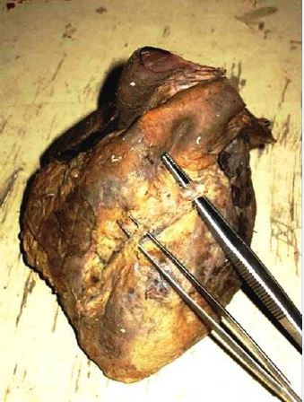
specimen was looked normal and free of any developmental defects. The examination revealed that there
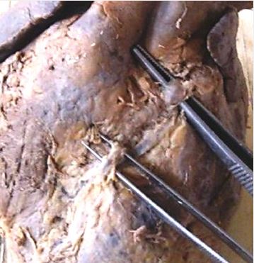
were two MBs; the first was on the middle third of the LAD (fig. 1), two bands were seen (fig. 2), the
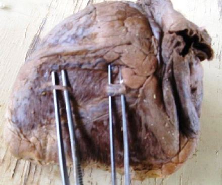
proximal (4mm) in length and the distal (12mm) in length, the second MB was on the posterior cardiac vein
(fig. 3); the MB was (17mm) in length and situated on the middle third of the vein. The both MBs were of
the superficial type.
Discussion
Myocardial bridging was first described in 1737, when Reyman noted this particular anomaly in human heart [3], it is an anatomical variant that affects the coronary arteries [1,2]. Multiple MBs were seen in the current case which agrees with Ferreira et al [4] and Cakmak et al [5]. Ferreira et al [4] reported two different types of myocardial bridges superficial type and deep type, in the current case only the superficial type was seen. The LAD artery is the most commonly affected artery [1,4]
The presence of MBs on the cardiac veins generally [6] and posterior cardiac vein specifically was not reported before the time of the present case since the MBs are related to the coronary arteries as general and LAD exclusively.
Conclusion
Myocardial bridging is a normal anatomical variant, generally a benign condition and common autopsy
finding. MBs easily seen in dissected specimens comparing to angiograms. It commonly localized to the
middle third of LAD branch of LCA. It rarely found in RCA and CCA. In the present case, an unusual
bridge was on the PCV.
Bibliography


Hi!
We're here to answer your questions!
Send us a message via Whatsapp, and we'll reply the moment we're available!