Biography
Interests
Emanso Umobong1, Ojo, B. A.2*, Ojo, O. N.3, Ikenna Okonkwo4, Mandus Akonjom5, Ugwu Ifeanyi6, Efu, E. M.7, Inemesit, D. E.8 & Eke Barnabass, A.9
1Histoconsult laboratory, Abuja, Nigeria
2Department of Anatomic Pathology, College of Health Sciences, Benue State University, Makurdi, Nigeria
3McMaster University, Hamilton Ontario, Canada
4Department of Pathology, Maitama District Hospital, Abuja, Nigeria
5Department of Surgery, State House Clinic, Abuja, Nigeria
6National Orthopedic Hospital, Enugu, Nigeria
7Department of Anesthesia and Intensive Care, Federal University Health Science, Oturkpo, Nigeria
8Screenics Pathology Solutions, Ondo, Nigeria
9Eke Barnabass A., Department of Surgry, College of Health Sciences, Benue State University, Makurdi, Nigeria
*Correspondence to: Dr. Ojo, B. A., Department of Anatomic Pathology, College of Health Sciences, Benue State University, Makurdi, Nigeria.
Copyright © 2023 Dr. Ojo, B. A., et al. This is an open access article distributed under the Creative Commons Attribution License, which permits unrestricted use, distribution, and reproduction in any medium, provided the original work is properly cited.
Abstract
A granular cell tumor (GCT) originating in the appendix is relatively rare and usually diagnosed as an incidental finding. Granular cell tumors are usually benign lesion of neural/schwannian origin, most frequently found in middle age women. Most patients were diagnosed preoperatively as having acute appendicitis. We describe the first case of GCT of the appendix in Abuja, Nigeria federal capital city in a 40 year old female patient who presented with signs and symptoms of appendicitis.
Introduction
Granular cell tumor (GCT) of the appendix is a rare tumor with only sixteen cases reported in over half a
century in the literature with no cases documented from Asia, Africa and Oceania and no cases of malignant
GCT of the appendix reported [1].
GCT was first described in the literature in 1854 as a cluster of large cells featuring granular eosinophilic cytoplasm [2] and named as granular cell myoblastomas in 1926 by Abrikosoff [3]. They are generally believed to originate from neural/schwannian cells, it can occur at any age, in any part of the body with peak incidence between 40 and 60 years of age and they most occur in women and black people [1,4,5-7].
We herein describe the first case of a GCT of the appendix in Abuja, the Federal Capital city of Nigeria with a detailed literature review.
Case Description
A 40 year old woman presented with a 2 day history of right iliac fossa pain and vomiting of 1 day duration, not
projectile. At physical examination, there was pain at the McBurney point radiating to the back. Abdominal
ultrasound revealed an edematous appendix with a traverse diameter of 7mm, peri- appendicular fluid
collection with thickened pouch and peri-appendicular fat. Given the clinical presentation and radiological
findings an acute appendicitis diagnoses was made and patient underwent appendicectomy. The surgical
specimen was fixed in 10% neutral formalin and consists of the vermiform appendix measuring 7.0cm
in length and 2.0cm at the widest diameter with dilated proximal end. The cut surface shows lumen with
facecloth. It was then paraffin embedded after surgical grossing.
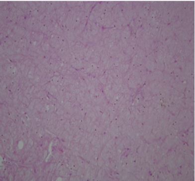
Besides haematoxylin-eosin (H&E) staining, immune-histochemistry (S-100) was performed. Specimen slides examination revealed with H.E, poorly defined sheets and nests of large cells separated by fibrocollagenous bands in the muscularis propria and the serosa. The cells have brightly eosinophillic coarse granular cytoplasm and small dense hyperchomatic nuclei and mild inflammatory infiltrates. No necrosis or mitosis seen (Fig. 1)
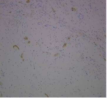
The tumor cells resulted immunoreactive for S-100 protein (Fig 2,). The post-operative phase was uneventful and was discharged home on postoperative day six and was seen a week after discharge
Discussion
Also called Abrikossoff tumor (3) GCT are rare type of soft tumor that usually begin in Schwann cells and
can occur anywhere in the body. One of its characteristics is that eosinophillic granules are contained in the
cytoplasm of cells [8].
The classic location of GCT is the tongue but it has been seen, however, in other locations such as skin, vulva, breast, larynx, bronchus, esophagus, stomach, appendix, rectum, anus, salivary glands, bile ducts, pancreas, urinary bladder, uterus, brain, pituitary gland, and soft tissue [9-18].
Congenital examples have been reported especially involving the gingival [19,20].
Most case of GCT is benign with malignancy occurring in 1% or 2% of cases [21,22]. From a histopathology perspective, Fanbury Smith and colleagues, [22] proposed the following criteria to determine whether a tumor is malignant or not: (i). The presence of necrosis (ii) the emergency of spindle cells, (iii) a vacuolar nucleus with an enlarged nuclear body (iv) increase in nuclear division (2 mitoses/10HPF), (v) increase in the nucleo-plasmic ratio and (vi) polymorphism.
If none of these diagnostic criteria are met, the tumor is considered to be benign. In one or two criteria are met, the tumor is considered to be atypical, and if three or more criteria are met, the tumor is considered to be malignant.
Immunohistochemically, positivity has been described for S-100 protein, positivity is also seen in laminin , calretinin, the alpha submit of inhibin HLA-DR, PGP 9.5, CD68, osteopontin, myelin basic protein, and CEA [8,23-28].
A GCT originating in the digestive tract, particularly in the appendix is relatively uncommon and very rare. They are usually diagnosed as an incidental finding. The index case was diagnosed after surgical pathology of an appendectomy specimen. She was a 40 year old black woman who presented with right iliac fossa pain. The peak incidences for GCT are between 40 and 60years and tend to occur in women and black people [1,4-7].
The surgical pathology for the index case described an appendix tissue cells having brightly eosinophilic coarse granular cytoplasm and small dense hyperchromatic nuclei and mild inflammatory. The S-100 protein was positive. In GCT, the cytoplasm is highly granular with most granules been small and regular. Immunohistochemisty positivity has been described for 5-100 among other proteins.
Conclusion
Granular cell tumor classically known as granular cell myoblastoma is uncommon and that of the appendix
is extremely rare. We described a benign case that was diagnosed as an incidental fining after surgical
pathology of an appendectomy specimen.
Consent
Written informed consent was obtained from the patient for publication of this report and any accompanying
images.
Findings
No extramural funding was used for this study.
Competing Interest
The authors declare no conflicts of interest with respect to authorship and/or publication of this article.
Bibliography


Hi!
We're here to answer your questions!
Send us a message via Whatsapp, and we'll reply the moment we're available!