Biography
Interests
Michel Leclerc
Immunology of Invertebrates, Orléans University, France
*Correspondence to: Dr. Michel Leclerc, Immunology of Invertebrates, Orléans University, France.
Copyright © 2022 Dr. Michel Leclerc. This is an open access article distributed under the Creative Commons Attribution License, which permits unrestricted use, distribution, and reproduction in any medium, provided the original work is properly cited.
Abstract
Sea star Axial organ (A.O) cells were observed in TEM. The axial organ has been described by us as a primitive lymphoïd organ in the years 1970. Sea star lymphocytes, platelets, have already been observed in previous works. They are smaller in diameter (4 to 5µ) than Vertebrate ones It seems to us that Thrombocytes exist also in this Invertebrate (TEM observations): Genomics asserts this last data.
Introduction
Observation of sea star Asterias rubens T and B lymphocytes have already been performed in TEM [1,2]: it
was asserted by biochemistry and biophysical assays. Second sea star platelets [3] were observed. Genomics
[3] assert these data.
In Vertebrates Hemostasis is a defence mechanism to prevent loss of blood in the event of an injury in an organism that has a vasculature [4]. In sea star (Upper Invertebrates) there is no blood « sensu stricto » but haemolymph (in the coelomic cavity). We try in the present work to analyse new images which seem correspond to Thrombocytes. and to aggregated platelets in transmission electron microscopy (TEM) at the level of the axial organ (AO).
Materials and Methods
Sea star Asterias rubens were obtained from the Marine Institute of Arcachon (France)
Axial organs (A.O) were excised and:
a) Either the whole cellular population was conserved and so observed in TEM
b) or the whole A.O was separated into B and T cell subpopulations according the well-known method of
Julius and al [5].
Cells were fixed with glutaraldehyde at 2%, in cacodylate buffer, as precedently described [1]
No post-coloration was operated.
Observations were done with a Hitachi Microscope.
Results
We have a look on the Figure below: the scale (in the bottom, right side) represents 1µ.
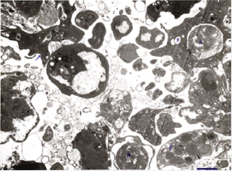
We observe always on this side what we descibed as a typical platelet (P) [3] without nucleus (N). Besides of it azurophiles particles smaller than 0,2µ are present.
These particles may correspond to lysosomes or to empty vesicles. Next this platelet we observe a nucleus (N) alone which is surrounded by a double membrane (nuclear membrane?): the nucleus is similar to the one which is shown on the top right side of the Figure. There, it is included in a platelet with digitations situated near another platelet: they seem aggregated.
We may envisage that the platelet with nucleus is an ancient thrombocyte which is « loosing » it. Note the large perinuclear space which is not due to a problem of fixation with glutaraldehyde but seems indicate this loosing.
Always in the top of the Figure, in the left side we observe another platelet (see arrow) with digitations. THERE IS NO NUCLEUS in this platelet.
Is it the definitive form of Platelet? In sea star system?
On a genomic point of view, it was demonstrated that thromboxane genes exist in this Invertebrate (Leclerc, 2019) [4].
Conclusion
The sea star Asterias rubens presents T and B lymphocytes, Monocytes, Macrophages and Platelets, in
T.E.M observations. The evolution of Thrombocytes in Platelets is envisaged from a point of view of
morphogenesis in Invertebrates. Platelets are seen with various aspects either with nucleus or without one.
Genomics confirms these data.
Nevertheless we have not yet observed classical Thrombocytes with nucleus showing a typical « indentation »as it is shown in human [4].
Bibliography


Hi!
We're here to answer your questions!
Send us a message via Whatsapp, and we'll reply the moment we're available!