CPQ Medicine (2018) 1:3
Case Report

Resected Adrenal Myelolipomas Discovered During Clinical Investigations for Abdominal Symptoms in Three Female Patients


Dr. Nasuhi Engin Aydin

Formerly at Department of Pathology, Izmir Katip Celebi University, Izmir, Turkey

*Correspondence to: Dr. Nasuhi Engin Aydin, c/o Pathology Laboratory, Ataturk Hospital, Izmir,Turkey.

Copyright © 2018 Dr. Nasuhi Engin Aydin. This is an open access article distributed under the Creative Commons Attribution License, which permits unrestricted use, distribution, and reproduction in any medium, provided the original work is properly cited.

Received: 25 April 2018
Published: 18 May 2018

Keywords: Myelolipoma; Adrenal Mass; Incidentaloma


Abstract

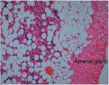
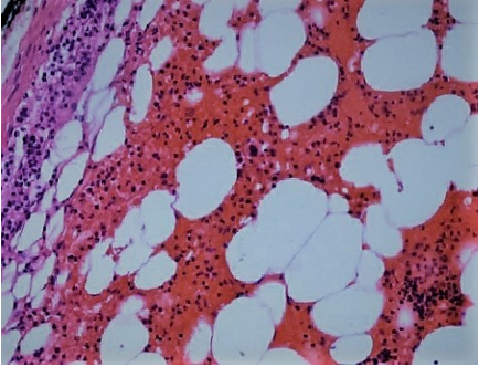
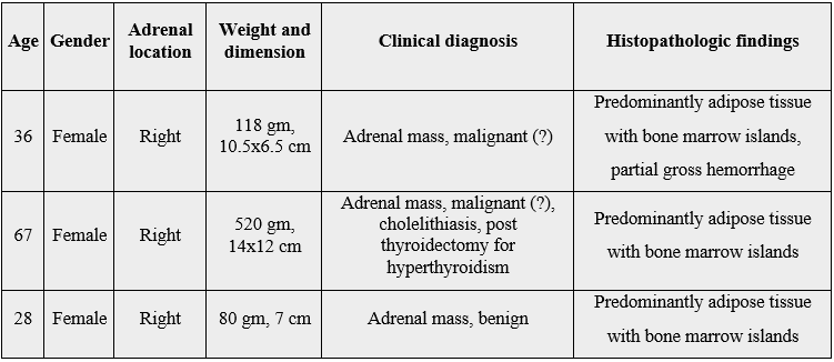
Adrenal myelolipomas are rare benign tumors composed of mature fat and hematopoietic tissue, commonly referred as the second most frequent “incidentaloma” of adrenals. Here three female patients with surgically resected myelolipomas all from the right adrenal are presented. The tissue was biggest in the elderly patient and all were predominantly rich in adipose tissue without any accompanying coincidental neoplasm. All the patients were in good health after a mean follow up period of six years.

Introduction
Myelolipomas (ML) is a rare benign tumor composed of mature fat and hematopoietic tissue with the most frequent location being the adrenal glands [1,2]. The earliest descriptions date back to the beginning of twentieth century and were termed as “formations myelolipomatoses” by Oberling in 1929. Thought to arise from metaplasia of undifferentiated stromal cells, these tumors used to be discovered primarily at autopsies; however, today they are typically incidental findings in living patients due to the widespread use of radiologic imaging techniques. Since these masses are asymptomatic and generally discovered unexpectedly they are also regarded as “incidentilomas” having a high prevalence after adrenal cortical adenomas [1,2].

Case Presentations
Case 1 A 36-year-old woman with complaints of abdominal discomfort was found to have high systolic blood pressure, 160/80mmHg and a 10x6cm right adrenal mass in US and MRI. Due to the large dimension of the mass a malignant adrenal neoplasm was also considered. Laparoscopic surgery was done with successful total excision of the mass. The specimen received at the pathology laboratory was 118gm, 10.5x6.5x6cm yellow mass showing predominantly adipose tissue with partially hemorrhagic appearance, (Figures 1 and 2). Hematopoietic tissue with immunohistochemically CD 33 and CD 61 positive cells among diffuse adipose tissue were seen in histologic sections (Figures 3 and 4). The patient was in good health without recurrence during follow up for six years.


Figure 1: Resected ML with partially encapsulated smooth border


Figure 2: Cut surface of the AML in Figure 1, showing diffuse yellow adipose tissue with brownish color in one pole


Figure 3: Low power microscopic view showing attenuated adrenal cortical tissue due to expansile fat with hematopoietic tissue (Hematoxylin and eosin x200)


Figure 4: Acute hemorrhagic hematopoietic tissue elements with megakaryocytes, (Hematoxylin and eosin x200)

Case 2 A 67-year-old woman who had total thyroidectomy for hyperthyroidism nine years ago presented with complaints of right upper quadrant pain and discomfort. She was found to have gallstones along with a large mass in the right adrenal following US and MRI. The patient was operated and the mass successfully resected along with cholecystectomy. She was hypotensive postoperatively and she recovered after three days in intensive care unit. The patient was in good health after follow up period of six years. Pathologic examination revealed a 520gm, 14x12x7cm yellow, nodular, adipose tissue mass. Hematopoietic tissue was seen among fat in microscopic sections yielding diagnosis of ML. The cholecystectomy revealed multiple gallstones and chronic inflammation. The patient was in good health during clinical follow up of six years.

Case 3 A 28-year-old woman was found to have a right adrenal mass during examination for abdominal discomfort. Laboratory tests were within normal limits including cortisol and ACTH levels. Laparoscopic surgical attempt for excision resulted in abdominal hemorrhage due to peripheral adhesions and the mass was resected totally by open surgery. Tissue received at pathology laboratory was 76gm, 7x6x5cm mass and microscopic sections showed benign adipose tissue along with islands of hematopoietic elements, i.e. ML. The patient became pregnant, gave birth to a healthy child and was in good health after a clinical follow up of seven years.

Discussion
Mass lesions unexpectedly discovered in adrenal glands are referred as “incidentiloma”. ML is the most common incidentiloma after adrenal adenoma and comprise approximately 3% of primary adrenal tumors [1-2]. They are more common in females between fifth and seventh decades of life and in the right adrenal gland. There is an increase in incidence of reporting this entity in the recent years, about 80% of the cases reported in the last 20 years and this is assumed to be due to the increased use of imaging modalities [1-8]. Myelolipomas can rarely appear at other sites of the human body, as well, hence the term extraadrenal ML [1,2]. Among numerous locations, myelolipomas have been described in the mediastinum, spleen, kidney, bones, thorax, nasal cavity, ectopic adrenal cortex, as well as at extradural sites or in the eyes [1,2]. Biochemically, myelolipomas are mostly non-functional. The mean maximum dimension of the adrenal myelolipoma ranges from 4 to 6 cms [1,2], huge adrenal MLs, i.e. bigger than 7cm, bear a risk of retroperitoneal hemorrhage [4]. One of the resected tumors among the present series also showed a 
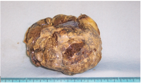
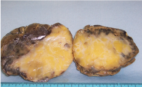
partially hemorrhagic appearance (Figures 1 and 2). The patient was operated before developing any clinical symptoms related to the aggravation of this complication. The three female patients with right adrenal mass lesions presented here were subject to surgery with successful outcomes without any recurrence after a follow up period of six and seven years. The two younger patients in this report had laparoscopic surgical approach with the results reported previously [6]. The older patient who also had gallstones directly underwent open surgery since the mass was bigger and cholecystectomy was done at the same time. However, she was hypotensive postoperatively necessitating for a three-day ICU treatment. This was possibly related to the metabolic status of the patient due to the previous thyroidectomy of nine years ago for hyperthyroidism. The association of ML with gallstones in this patient seemed coincidental but there is an element of suspicion regarding a common pathophysiologic basis [5].

There are cases of malignancy coincident with adrenal ML which necessitates meticilous pathologic examination [7,8]. In this regard, it is advisable to have a quick frozen section pathologic assessment intraoperatively to be certain about the nature of a lipomatous mass that seems to be benign. None of the three cases reported here had frozen section despite the fact that in two patients’ presumptive clinical diagnosis included malignancy due to the large dimensions noted.

Table: Features of the patients presented.


Bibliography

  1. Lam, A. K. (2017). Update on adrenal tumours in 2017 World Health Organization (WHO) of Endocrine Tumours. Endocr. Pathol., 28(3), 213-227.
  2. Decmann, A., Perge, P., Toth, M. & Igaz, P. (2018). Adrenal myelolipoma: a comprehensive review. Endocrine, 59(1), 7-15.
  3. D'Addosio, R., Rojas, J., Bermúdez. V., Ledesma, F. & Hoedebecke, K. (2017). An incidentaloma that catches your eye - adrenal myelolipoma. F1000Research, 6(1140).
  4. Wu, M. Y, Hou, Y. T. & Yiang, G. T. (2018). Acute retroperitoneal hemorrhage induced by giant adrenal myelolipoma mimicking renal colic pain. Reports, 1(1), 4.
  5. Bano, S., Yadav, S. N., Chaudhary, V. & Garga, U. C. (2012). Symptomatic giant adrenal myelolipoma associated with cholelithiasis: two case reports. Urol Ann., 4(1), 55-60.
  6. Ates, M., Karaca, Z., Dirican, A., Hatipoglu, S., Sahin, I. & Aydin, N. E. (2012). Laparascopy in adrenal myelolipoma. Med Science., 1(3), 200-5.
  7. Wang, J., Fisher, C. & Thway, K. (2014). Dominant myelolipoma encasing adrenal cortical carcinoma. Int J of Surg Path., 22(8), 731-735.
  8. Hagspiel, K. (2005). Manifestation of Hodgkin’s lymphoma in an adrenal myelolipoma. Eur Radiol., 15(8), 1757-1759.

Total Articles Published

8
9
2


Total Citations:

1
8
4


Highlights


Cient Periodique is a ‘Gold’ open access publisher that aspires to offer absolute free, unrestricted access to the valuable research information

We welcome all the eminent authors to submit your valuable paper

Cient Periodique invites the participation of honourable Editors and Authors

CPQ Journals provide Certificates for publication

Cient Periodique also offers memberships for potential Authors

Best Articles will be appreciated with the provision of corresponding Certificate

Hi!

We're here to answer your questions!


Send us a message via Whatsapp, and we'll reply the moment we're available!