Biography
Interests
Viren Shirish Patil*, Mayur Limbhore, Ramanojam Shandilya, Ajinkya Kadam & Shivani Chavan
Department of Oral and Maxillofacial Surgery, Bharati Vidyapeeth Dental College and Hospital, Pune, India
*Correspondence to: Dr. Viren Shirish Patil, Department of Oral and Maxillofacial Surgery, Bharati Vidyapeeth Dental College and Hospital, Pune, India.
Copyright © 2018 Dr. Viren Shirish Patil, et al. This is an open access article distributed under the Creative Commons Attribution License, which permits unrestricted use, distribution, and reproduction in any medium, provided the original work is properly cited.
Abstract
This paper reports a case of a recurrent periapical lesion treated with enucleation of the lesion, apicoectomy, and root end obturation on a lower anterior teeth and right first molar. In the case of conventional root canal treatment failure, non-surgical retreatment is the preferred option in most of the cases. Several factors such as a complex root canal system or previous procedural accidents may impede the success of non-surgical retreatment. The authors have performed root-end resection and preparation under local anesthesia on a lower left first molar and lower anterior teeth. The root canal filling is placed within the new cavity to close the path of communication between the infected root canal system and periradicular structures with an intermediate restorative material. The lesion was fully enucleated. The 49 year-old male patient was followed up and presenting as a functional and symptomless tooth. Radiographic findings showed a clear and progressive refilling of the cavity with bone. All these factors highlight a positive prognosis for the tooth after periradicular surgery, which is now considered a valid treatment to keep the tooth as a functional unit in the oral cavity.
Case 1
A 49 year-old male patient was referred by her dentist to our Oral Surgery Department for the extraction of
the lower right first molar (46). Her referral letter clearly explained the previous attempts for preserving the
tooth with endodontic treatment and a enucleation in the periapical area under local anesthesia.
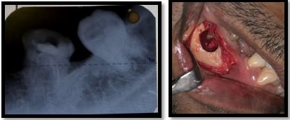
The mild anxious patient revealed her chief complaint as a “throbbing pain” in the right mandibular region which he defined as intermittent and spontaneous, but was also evoked by percussion. His second complaint was painful to touch intermittent swelling in the buccal aspect immediately below the gingival sulcus of the mandibular right posterior region. He reported that his dentist tried to restore the tooth with endodontic treatment and also performed an operation with local anesthesia. A periapical lesion was removed related to the apices of madibular posterior region (46) as included in the referral letter. The dentist’s attempts to preserve the tooth had limited success because he kept feeling pain. A periapical X-ray demonstrated persistent bone resorption. She had recurrent infections which needed to be treated with antibiotics. No sensory loss or numb/tingling sensation had ever been reported by the patient. Medical history: The patient is fit and well; the patient denied any major systemic disease. Low/mild dental anxiety levels as reported by the pre-assessment questionnaire, but the patient was amendable to local anesthesia and already has undergone prolonged dental procedures under local anesthesia. In an attachment to the dentist’s letter was a small periapical X-ray (Figure 1) of mandibular posterior teeth (46) was attached. Considering the case, the surgeon and the clinic’s supervisor suggested to the patient to undergo an apicoectomy to preserve the tooth potentially. The X-ray examination showed a restorative filling and a well-done root canal filling on the 46 region with a periapical radiolucency (1cm ). The periapical lesion is in intimate contact with the inferior alveolar nerve canal (Figure 1).
On the day of the surgery, the patient was admitted to the clinic, the surgical procedures were described, the risks and benefits were explained, and a consent form was signed. The patient sat comfortably on the chair and asked to complete one preoperative mouthrinse with betadine. The sterile surgical tray was opened, sterile drapes were used to cover the patient. Local anesthesia was performed using 2 cartridges of Lignocaine (2% lidocaine with 1:2,00,000 epinephrine injection solution) 1 cartridge for the lower right inferior alveolar block plus 1 cartridges used for wide buccal infiltration from lower right 45 to 48 teeth region.
According to the classic literature, the most commonly used mucoperiosteal flaps for apical access in the
posterior part of the mandible are the envelope flap and the triangular flap with an obvious mesial releasing
incision. The releasing incision preserves the distal vascular branches from the buccal artery. Considering
the proximity to the mental foramen, we elected to choose a different surgical approach that allowed
us to gain better access to the mandibular posterior root region. This surgical approach increases visibility,
reduces the amount of exposed bone, and reduces the risk of mental nerve damage. The supervisor and the
first operating surgeon proposed this mucoperiosteal flap with a wide angled distal relieving incision.
After elevating the flap and visualizing the cortical bone, immediately a continuous soft tissue structure
was noted between the bony cavity and the buccal flap. After dissection, the buccal bone showed a hollow
space slightly mesial to the 46. Ostectomy was performed successfully with a round carbide bur with the
opportunity to enlarge the bony defect to a buccal window (1cm ) and gaining access to the cystic structure
and the 46 roots (Figure 2).
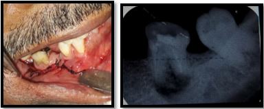
The process of removal of the periapical lesion was incomplete in the lower part of the cavity. In fact, because of the proximity of the alveolar nerve, curettage provoked an intense sensation while attempting to elevate the bottom of the periapical cystic surrounding fibrous tissue. This discomfort led the surgeon to choose a more conservative approach during the operation.
A straight fissure carbide bur was directed perpendicular to the long axis of each root and used with full rotating power to cut the mesial and distal 3mm of the apex. This cut is completed in order to decrease dentin tubules peripheral microfiltration. This cut exposed the gutta percha material and removed any excess gutta percha protruding from the apical foramen. Saline irrigation was used to clean the cavity, removing debris of gutta-percha, dentin, and cementum (30s). Sterile gauzes in long strips were gently placed inside the empty cavity to obtain hemostasis, to clean the environment. The strips were removed after 2min. The 2 apices of the roots were visualized and then carefully resected at 90° with a straight fissure bur on a handpiece]. The careful use of a manual bone trimmer allowed us to plane and remove any blunt border from the window access. The trimming was completed to enhance soft tissue juxtaposition and healing. Before closing the flap, the removal of periradicular fibrous tissue was performed with the hope of gaining a thinner, less prone to inflammation gingiva, even if the clinical outcome would have shown a slight recession of the gingival margin. The area has no esthetic consideration, so the patient was satisfied (Figure 3).
Case 2
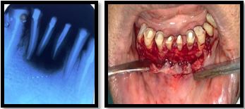
A 60 years old male patient reported to department of oral and maxillofacial surgery with a chief complaint
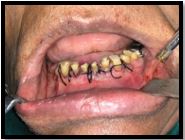
of perapical lesion over mandibular anterior region since 15 to 20 days. An intra oral periapical radiograph
was advice to the patient which revealed periapical lesion with respect to Root canal treated teeth that is
mandibular right and left central incisior (41,31) and right lateral incisor (42) region (Figure 1).
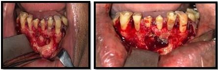
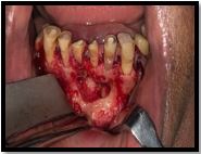
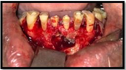
Enuclection and Surgical removal of periapical lesion followed by apicoectomy was planned under Local anesthesia. Painting and draping was done following all aseptic precautation. Lignocaine with adrenaline (1: 2,00,000) was infiltrated at the surgical site. Crevicular incision was taken. (Figure 2). Mucoperiosteal flap was raised. Periapical lesion seems to invade the overlying bone. Bone window was created with the help of round bur and periapical lesion was exposed (Figure 3). Enucleation of periapical cyst was carried out.
Apicoectomy of 31,41,42 was carried out with help of straight fissure bur (3mm) and excess of gutta percha points were removed. Apical sealing was done with help of mineral tri oxide (Figure 4) and cavity was irrigated with help of normal saline and betadine. Suturing was done. (Figure 5) Follow up of patient was taken for 3, 7, 10 days.

Discussion
After the failure of the conventional root canal treatment (RCT), non-surgical retreatment is the preferred
option in most cases. Several factors, such as a complex root canal system or previous procedural accidents,
may impede the success of non-surgical retreatment. In these cases, periradicular surgery and apicoectomy
would be the treatment of choice to preserve the tooth.
Performed root-end resection and preparation, the root canal filling is placed within the created cavity to close the path of communication between infected root canal system and periradicular tissues. Using a root canal filling material with ideal properties has an immense effect on the treatment outcome of the surgery. An ideal material should be non-absorbable, non-corrosive, non-cytotoxic, not affected by moisture, dimensionally stable, biocompatible, antibacterial, radiopaque, cost-effective, easily manipulated, adhesive to dentinal walls, create a tight seal, and induce cementogenesis [1].
Numerous materials have been recommended for root-end obturation, and many studies have attempted to identify an ideal material; however, an ideal material has not been found [2]. The most commonly used materials are mineral trioxide aggregate (MTA) and IRM [2]. The follow-up examinations of this cases were after 2 days. The examinations noted an absence of symptoms, such as swelling, trismus and the normal function of the tooth was preserved. No sensory loss was reported by the patient.
The patient is happy and satisfied to have her tooth functional. Also, the X-ray evaluation reported clear and slow bone deposition in the cavity after apicoectomy, obturation, and cystic enucleation. These criteria highlight the success of the treatment after 6 months. Therefore, after the previous failure of the endodontic treatment, we can consider this a valid treatment.
This is a bold act, with a high rate of success that must be considered on certain occasions by the experienced surgeon. This procedure was suggested to the patient as an act of pure preservation and to respect the anatomo-functional importance of the tooth [3].
Conclusion
Based on the contemporary understanding of apiceoctomy procedure for success and failure, assessment
and subsequent treatment have greatly improved. Advances in apiceoctomy armamentaria and materials
(including retrofilling materials and clinical techniques for their efficient use) have enabled Oral and
Maxillofacial to treat challenging cases with much greater efficiency.
Conflicts of Interest
There are no conflicts of interest.
Patient Consent
Obtained.
Bibliography


Hi!
We're here to answer your questions!
Send us a message via Whatsapp, and we'll reply the moment we're available!