Biography
Interests
Pani Jyoti Prakash1*, Philip Chandra, X.1, Gupta Rashmi2, Pani Sankarsan3, Singh Royana4
1Department of Anatomy Mahatma Gandhi Mission Medical College & Research Institute, Pilliarkuppam,
Pondicherry
2Department of Bioinformatics University of North Dakota, United States of America, Post Doctoral Fellowship
stream
3Progressive Universal Society, Bharatpur, Rajasthan, Jaipur, India
4Department of Anatomy Institute of Medical Sciences Banaras Hindu University Varanasi, Uttar Pradesh
*Correspondence to: Dr. Pani Jyoti Prakash, Department of Anatomy Mahatma Gandhi Mission Medical College & Research Institute, Pilliarkuppam, Pondicherry.
Copyright © 2019 Dr. Pani Jyoti Prakash, et al. This is an open access article distributed under the Creative Commons Attribution License, which permits unrestricted use, distribution, and reproduction in any medium, provided the original work is properly cited.
Abstract
The bad impact of daily prolonged administration of silver nanoparticle colloidal solution on cognitive function of Pregnant Swiss Albino mice and their fresh born fetuses were studied. The over building up of nano metallic silver in whole hippocampus tissue and residual part of mice brain was investigated by keane observation of high precise and representative psychic state and its analysis and the meticulous review of histological study were conducted in the present research. Some of the main result of this comprehensive study is description of hippocampus. Stockpiling of nano metallic silver in the pregnant mice mature hippocampus and fetal developing hippocampus inside brain lead to integrity violation of the CA1, CA2, CA3 and CA4 region caused long term contextual memory impairment with visible changes in psychic behavior and cognitive hindrance. This leads to altered psychic state and psychosomatic disorder. Usually 3 types of sizes of nano silver metal are available in market extreme small size (0.25 to 1 nm sizes ranges), moderate small or medium size(1.25 to 20.38nm sizes ranges) and larger size(20.75 to 100.55nm sizes ranges) where sizes were focused on the external aspect of package after releasing from manufacturing company. The present study aims to investigate the effect of prenatal exposure to small and large size AgNps on cognition and altered hippocampus response. We commenced serial daily morning (9AM) once repeated oral gavages ingestion experiment with magnetic stirring and cooling fresh made AgNps solution of above 3 sizes on pregnant Swiss Albino mice and their freshly born fetuses (10 from each group treated and sham control and from 6 to 19 gestational days(pregnant mother) and 1month (fetuses after delivery)) and found both sufferers of the same (Toxic and teratogenic hippocampus response which is directly dependant on size and dose) after nano silver metal crosses blood brain barrier reaching hippocampus and held inside such white matter tissue. These nano silver metallic particles also crosses embryonic membrane reaches fetus brain in mother uterus and hoarding in developing hippocampus due to same mechanism. These various admixture nano silver particles also surprisingly demonstrated gender specific stinging exhibition. Our scientific expert panel have collected and assessed the corrosive and hazardous sign and symptoms of 14 Gestational days(6 to 19 gestational days (pregnant mother) and 1 month (fetuses after delivery)) mature and developing hippocampus response with once daily continuous mouth ingestion of volumetric AgNps fresh solution having admixture of 0.27 to 1.03nm size range (smallest), 1.28 to 20.41nm size ranges(smaller medium) & 20.78 to 100.53nm size ranges (larger) at a lowest dose of 0.37(1) , 0.56(2) , 0.73(3)mg/kg b.w. in an in between dose of 4.2(1), 8.2(2) & 12.2(3)mg/ kg b.w. (moderate) and in a higher dose 16.1(1), 20.1(2) & 24.1(3)mg/kg b.w. (larger). Groups were assigned as 1(Sham Control); 2(2a,2b,2c), 3(3a,3b,3c) & 4(4a,4b,4c) (Treated). At lowest dose (given to each 30 mother and fetuses) we report minimal altered and toxic hippocampus histological findings, the moderate doses showed in between teratogenic and toxic result and at larger doses our panel reported predominant hippocampus histological teratogenic as well as toxic and altered psychic irritative findings. These above mentioned size and dose range group nano metallic silver treatment leads to atrophic and degenerated hippocampus proprius (CA1, CA3 and CA4) along with atrophied, degenerated and distorted molecular, pyramidal and granular cell layer also injured, break and weak axons and dendrites were found in histological panoramic view. (10X and 400X magnification of binocular bright field microscope) The moderate and higher dose showed admixture of teratogenic and toxic consequences whereas lower dose and size showed only toxic sign and symptoms. Maternal exposure to polyvinyl pyrollidone coated and sodium borohydried stabilized metallic nano silver in colloidal form during pregnancy hampered balanced cognition and psychic state in pregnant mice and offspring’s. Cognitive impairment purely dependant on one specific protein encoded gene depletion which secretes from nerve ending and reduce neurite outgrowth. The hippocampus toxicity and teratogenicity was mainly associated with disbursement of ionic silver.
Introduction
In this present and past era of nanotechnology globalization the first line consumer appraised metallic nano
research and market product is nano form of silver metal. But the raw metal changes to osmolality solution,
powder or transparent crystal form after biological, herbal and chemical coalescence. After successful
portraiture different sizes and form of these nano metal calculated if admixture also segregated and tagged
to various sizes ranges. Then in the nano research laboratory these various form of silver metallic nano were
confirmed to various larger and smaller size ranges by confocal microscopy and scanning electron microscopy.
After requisite laboratory processing individual size from smaller towards larger in safe packing delivered
to market for selling which are utilized for research purpose and company manufacture of various patent
consumer products for business purpose. From the initiation of 2000 era nano metallic silver have been in
utilized in several applications in medicament stream. [1-4] also it is peakly utilized in fabric industries [5-
7]. In recent years we also see heavy use of metallic nano silver in aseptic consumer products [8], now a day’s
also it is highly accepted to be utilized as rappers and foils for packing solid food stuffs [9]. Metallic nano
silver also highly accepted for additives in regularly intake foods [10-14]. All these because nano Ag possess
specific bactericidal and disinfectant properties that were well recognized since ancient past [15]. For this
reason only publics of old Raja maharaja times successfully used silver utensils as hygienic [16]. Still up to
today nano metallic silver competes to become successful drug coat for new generation antibiotics [17,18].
Obviously question evokes in the brain of scientists that why for we not accepts to visible silver macro form and but yes to silver of nano form. The answer for this is; nano form is accepted by the eco and aqua nature which produces likeliness to it also produces certain hazardous effect. From the beginning of the earth Darwinism or natural selection nature restructure techniques to counteract with natural nano sized silver metallic filaggrin and inter cellular structures. For example a livelihood nourishment compartment can acknowledge filaggrin in a indigenous or domestic situation [19]. These domestic situation are always maintain size of a silver nanoparticle as an engulfing agent like fast eating of solid food or drinking of liquid food. For this reason only particle colloid metallic nano silver are regarded as engineered nanoparticle either a layer of coronal filaggrin is there or not considered as nourishment or building material for the lively chamber. The technique of engulfing such as bodily process [20] and microendocytosis [21] is done by nano sized developed silver metallic material in this atmosphere. This type of activity leads to strong penetration of actively engineered silver nano metallic particle inside the cell and their accumulation and bioactivity inside into cellular and tissue space. For example nano metallic silver can assemble in vital neural organs such as different part of the brain and especially in hippocampus region of Pregnant mammals and their freshly born offspring’s [22-25]. Several researches showed brain especially hippocampus tissue is prone for prolong and critical trapping of silver nanoparticles [23,24] and some nano scientific report confidently explains riddling of small size nano metallic silver through handrail or crash barrier and their tightly holding in the hippocampus tissue [23,24,26]. Therefore hippocampus can be considered as the peak point goal organ for small size also for large and medium size metallic nano silver. At the same time those size metallic nano silver makes a chemical interaction with the hippocampus cells and proceeds to morphological cell damage. Earlier it was assessed that accumulations of those size nano metallic silver cause hippocampus cells disintegrate position and damaging and degeneration of the crash barrier. This happens due to speed illumination of such particles from hippocampus tissue in 4 month of intra uterine life and AgNps accumulation.
Most of the scientific researches demonstrate toxicity of metallic nano silver to Neurons, Astrocytes and Glial cells of hippocampus tissue [27-35]. The intensity of toxicity and teratogenicity depends directly upon dose, size and experimental exposure period.
Nano metallic silver when overwhelmed in intra tissue and cell space of hippocampus it produces cumulative toxicity in that region also some nano scientific researches give emphasis to psychiatric or sublimation and etiquette function of model mammal like mice and rat to see the influence of toxicity after accumulation of AgNps in hippocampus tissue [36-41]. Usually such researches are conducted for no more than 30 days and the doses of AgNps are able to poison animals without acting on the hippocampus [42].
Excessive use of silver nanoparticles commences in the field of medicine due to strong antibacterial and anti fungal activity [43]. also it is used both in consumer products and medical products. This metallic nano silver also utilized for multiple therapeutic purposes such as burn wound and some other skin diseases. This nano metallic silver also utilized in heavy volume for manufacture of fast food wrapper, medicine transport media, cosmetics, filters, toys, shampoo and tooth paste. Such peak and excessive use of nano metallic silver causes hazard to human hippocampus region and brain nervous tissue. Many in vivo and in vitro researches shown the teratogenic and toxic disambiguated effects on hippocampus and on human brain in past [44,45]. The chronic diseases may arose in the body by prolong dermal contact and smelling of nano silver metallic particles [46]. The small or large size to volume ratio can undergo a series of hazards by small size nano metallic silver penetrate through cell and plasma membrane and cause chemical reaction with Deoxy Ribo Nucleic acid and cell filaggrin cause decease of cell and organ damage such as spleen , liver and kidney failure. Also cross the blood brain barrier to cause hippocampus toxicity where large sizes cause teratogenicity [47].
The aim of the present study was the complete exploration of the facts of the impact of low concentration PVP coated and NaBH4 stabilized nano metallic silver particle on pregnant mammal and their fetuses cognitive function, effect of held of quantity of nano metallic silver in disambiguated hippocampus and its morphological and histological altered studies from brain slices and slides. This phase of research study has been conducted with a proposal of repeated daily once oral gavages administration of nano metallic silver colloid fresh prepared solution with the use of ultra modern top quality visual equipment and magnetic stirring and cooling method with post hoc and P-value analysis.
Material & Method
We commenced serial daily morning (9AM) once repeated oral gavages ingestion experiment with
magnetic stirring and cooling fresh made AgNps solution of above 3 sizes on pregnant Swiss Albino mice
and their freshly born fetuses (10 from each group treated and sham control and from 6 to 19 gestational
days (pregnant mother) and 1 month (fetuses after delivery) and found both sufferers of the same (Toxic
and teratogenic hippocampus response which is directly dependant on size and dose) after nano silver metal
crosses blood brain barrier reaching hippocampus and held inside such white matter tissue. These nano
silver metallic particles also crosses embryonic membrane reaches fetus brain in mother uterus and hoarding
in developing hippocampus due to same mechanism. These various admixture nano silver particles also
surprisingly demonstrated gender specific stinging exhibition. Our scientific expert panel have collected
and assessed the corrosive and hazardous sign and symptoms of 14 Gestational days (6 to 19 gestational
days) (mother) and 1 month to fetuses after delivery for developing and mature hippocampus response
with once daily continuous mouth ingestion of volumetric AgNps fresh solution having admixture of 0.27
to 1.03nm size range (smallest), 1.28 to 20.41nm size ranges(smaller medium) & 20.78 to 100.53nm size
ranges (larger) at a lowest dose of 0.37(1) , 0.56(2) , 0.73(3)mg/kg b.w. in an in between dose of 4.2(1), 8.2(2)
& 12.2(3)mg/kg b.w. (moderate) and in a higher dose 16.1(1), 20.1(2) & 24.1(3)mg/kg b.w. (larger). Groups
were assigned as 1(Sham Control); 2(2a,2b,2c), 3(3a,3b,3c) & 4(4a,4b,4c) (Treated).
The study was carried out in strict conformity with laws and regulation for animal experiment after getting
approval from central animal ethical committee of the institute (No. Dean/ 2014/CAEC/614/Dt.30.05.14).
The study was conducted in strict accordance with the rules and regulation and recommendation in the
guide for the care and use of laboratory animals. Central Animal Ethical Committee, Division IMS, BHU
provided ethical clearance for this project also the recommendation.
Silver nitrate (AgNo3) of 98.3% pure and transparent with shinning crystal having above mention three
sizes nano metallic silver purchased in 3 phases and admixture in a aseptic gamma ray radiated experimental
bottle and top sealing with a glass lead with proper leveling outside the date of admixture and mention of
the sizes sended to laboratory for preparation of magnetic stirring and cooling freshly prepared colloidal
solution. Crystal powder was purchased from Trimurthy Scientific world Varanasi. Solution was prepared
by double deionized water collected from distillation plant with addition of minimal volume Poly Vinyl
Pyrollidone (PVP) deaggregator (0.3% PVP solution: 0.1g of PVP was dissolved into 33ml distilled water)
and 0.002M NABh4 (Sodium Borohydried stabilizer/0.0189g of NaBH4 was dissolved into 250ml distilled
water.) and 0.001M AgNO3: 0.017g of AgNO3 was dissolved into 100ml distilled water. This solution can
be kept for later usage.
Magnetic stirring and cooling chemical reduction methods was used for synthesis of above mention sizes
nano metallic silver(0.27 to 1.03nm size range (smallest), 1.28 to 20.41nm size ranges(smaller medium) &
20.78 to 100.53nm size ranges (larger)) by using PVP (Poly Vinyl Pyrollidone and Sodium Borohydried
(NaBh4) deaggrgator, reducer and stabilizer agent. 0.001M of AgNo3 was cooled kept in a fresh burrate.
2ml AgNo3 solution was added drop by drop with the help of a switch guided dropper to 30ml NaBh4 fresh
solution finally 2 drops of PVP fresh solution was added to the existing. Over magnetic stirrer at 380Hz
for 20 minutes the solution was kept and stirred and final experimental solution was prepared. Cooling was
done by peri circumferential ice berg and mixed until color changed (Amber color). Then it removed from
magnetic stirred device (1 inch magnetic stirrer) and cooled to freeze 4˚c temperature and preserved for
application in experiment.
Pregnant Swiss Albino mice (25 to 31 weeks) and their freshly born fetuses (10 from each group treated
and sham control and from 6 to 19 gestational days treated with deionized double distilled water and with
once daily continuous mouth ingestion of volumetric AgNps fresh solution having admixture of 0.27 to
1.03nm size range (smallest), 1.28 to 20.41nm size ranges(smaller medium) & 20.78 to 100.53nm size
ranges (larger) at a lowest dose of 0.37(1) , 0.56(2) , 0.73(3)mg/kg b.w. in an in between dose of 4.2(1), 8.2(2)
& 12.2(3)mg/kg b.w. (moderate) and in a higher dose 16.1(1), 20.1(2) & 24.1(3)mg/kg b.w. (larger).
Group 1/ mice and fetuses served as sham control injected 0.25 ml and 0.125ml/ mice deionized double distilled water and was also injected for 6 to 19 gestational days (mother) and 1month(to fetuses after delivery).
Group 2(2a,2b,2c), 3(3a,3b,3c) & 4(4a,4b,4c) / mice and fetuses served as treated injected lowest dose of 0.37(1) , 0.56(2) , 0.73(3)mg/kg b.w. in an in between dose of 4.2(1), 8.2(2) & 12.2(3)mg/kg b.w. (moderate) and in a higher dose 16.1(1), 20.1(2) & 24.1(3)mg/kg b.w. (larger) and was also injected for 6 to 19 gestational days (mother) and 1month m(to fetuses after delivery).
After finishing oral gavages schedule to mother and fetuses at end the pregnant mice and 1 month developing
fetuses were sacrificed periodically and the brain tissue were separated. The tissue were fixed into 37 to 41%
formaldehyde solution in order to conduct histopathology experiment of hippocampus tissue, histological
sections were prepared from hippocampus and examined under bright field and light binocular microscope.
In between the experiment pregnant mother and fetuses’ cognition and behaviors are also meticulously
assessed.
The normal and altered histological and behavioral data were analyzed by multi ANOVA, Post hoc analysis
and P value analysis and comparison was made between control and test data.
Result
Some of the main result of this comprehensive study is description of hippocampus. Stockpiling of nano
metallic silver in the pregnant mice mature hippocampus and fetal developing hippocampus inside brain
lead to integrity violation of the CA1, CA2, CA3 and CA4 region caused long term contextual memory
impairment with visible changes in psychic behavior and cognitive hindrance. Which leads to altered psychic
state and psychosomatic disorder?
Toxic and teratogenic hippocampus response which is directly dependant on size and dose after nano silver metal crosses blood brain barrier reaching hippocampus and held inside such white matter tissue. These nano silver metallic particles also crosses embryonic membrane reaches fetus brain in mother uterus and hoarding in developing hippocampus due to same mechanism. These various admixture nano silver particles also surprisingly demonstrated gender specific stinging exhibition.
At lowest dose (given to each treated group 30 mother and fetuses) we report minimal altered and toxic hippocampus histological findings, the moderate doses showed in between teratogenic and toxic result and at larger doses our panel reported predominant hippocampus histological teratogenic as well as toxic and altered psychic irritative findings. These above mentioned size and dose range group nanometallic silver treatment leads to atrophic and degenerated hippocampus proprius (CA1, CA3 and CA4) along with atrophied, degenerated and distorted molecular, pyramidal and granular cell layer also injured, break and weak axons and dendrites were found in histological panoramic view. (10X and 400X magnification of binocular bright field microscope) The moderate and higher dose showed admixture of teratogenic and toxic consequences whereas lower dose and size showed only toxic sign and symptoms.
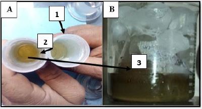
Chemical reduction with magnetic stirring and cooling method has used in this study for synthesis of
various sizes admixture metallic nano silver in colloidal form. The light ash color solution turned to Yellow
first then Amber color which is indicated synthesis of nano metallic silver. (Fig.1)
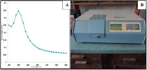
SL 164 Double Beam Uv-Vis Spectrophotometer (ELICO) was used to determine the surface Plasmon
resonance (SPR) bands,, from fig (2) the SPR bands detected around 420nm with absorbance 0.36. Spherical
(larger) and irregular multi spiny (smaller) shape of nanometallic silver contributed to the absorption band
around 600nm. The spherical and irregular multi spiny shape has been confirmed by transmission electron
microscopy.
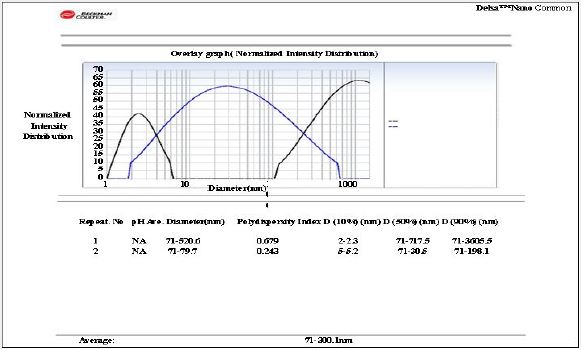
We estimated the admixture AgNps colloidal stirring solution sizes by overlay graph (Normalized Intensity Distribution) which is calculated by Delsa TM Nano common Beckman and Coulter DLS machine purchased from London; United Kingdom with the help of IIT, Pharmaceutical Division; Banaras Hindu University; Varanasi under the supervision of esteemed Professor Dr Sanjay Singh. We found admixture sizes are smallest (0.27 to 1.03nm size range), smaller medium (1.28 to 20.41nm size range) and larger (20.78 to 100.53nm size range). The above Overlay (Normalized Intensity Distribution) graph indicated almost 43 Hz Normalized Intensity Distribution peak in smallest size, 60Hz Normalized Intensity Distribution peak in smaller medium size and almost 65 HZ Normalized Intensity Distribution peak in larger size. Around avg. 0.461Poly Dispersity Index calculated for this type of stirring admixture sizes AgNps solution. 90% Density calculated was 71nm to 300.1 nm. So we came to a conclusion that maximum nanometallic silver particle size varies between 71nm to 300.1nm with avg. Diameter of 79.7nm.
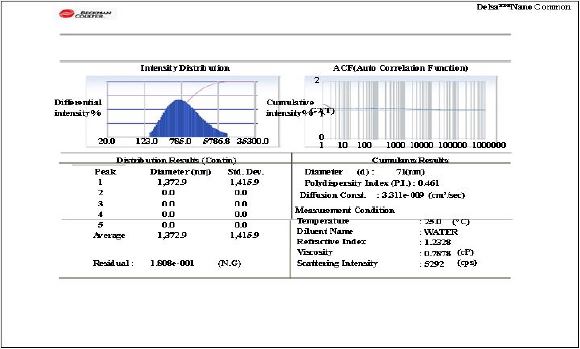
We also estimated the admixture AgNps colloidal stirring solution sizes by Intensity Distribution graph (Auto Correlation Function) which is calculated by Delsa TM Nano common Beckman and Coulter DLS machine purchased from London; United Kingdom with the help of IIT, Pharmaceutical Division; Banaras Hindu University; Varanasi under the supervision of esteemed Professor Dr Sanjay Singh. We found Differential Intensity Distribution result with the help of 5 peaks of diameters and its standard deviation. The avg. size was found 0 to 1372.9nm size ranges with a residual of 1.808e-001(N.G.). PDI was observed 0.461, Avg. Diam. Was observed 71nm, Diffusion constant was observed 3.331e-0.009, Viscosity of the solution was observed 0.7878cP and Scattering Intensity was observed 5292 cps. So we came to a conclusion that this AgNps stirring fresh cool colloidal solution possesses an admixture of smallest, smaller medium and larger sizes with an avg. diameter of 71.9 to 300.1nm size ranges of nano metallic silver.
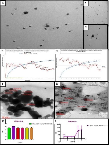
We also did TEM estimation (1.5 lakhs Magnification 50nm thick smear) of the admixture AgNps colloidal stirring solution for assessment of sizes of nano metallic silver with the help of IITR (Indian Institute of Toxicological Research; Luck now) Transmission Electron Microscopy Division; Luck now University; Luck now under the supervision of esteemed Professor Dr L. C. Chauhan. Whole TEM plots are photographed from 50nm thick smear. In this we found three categories of sizes of nano metallic silver. Smallest, smaller medium and larger. Smallest indicate 0.27 to 1.03nm size range, smaller medium indicate 1.28 to 20.41nm size range and larger indicate 20.78 to 100.53nm size range. So we came to a conclusion that this AgNps stirring fresh cool colloidal solution possesses an admixture of smallest, smaller medium and larger sizes with an avg. diameter of 71.9 to 300.1nm size ranges of nano metallic silver.
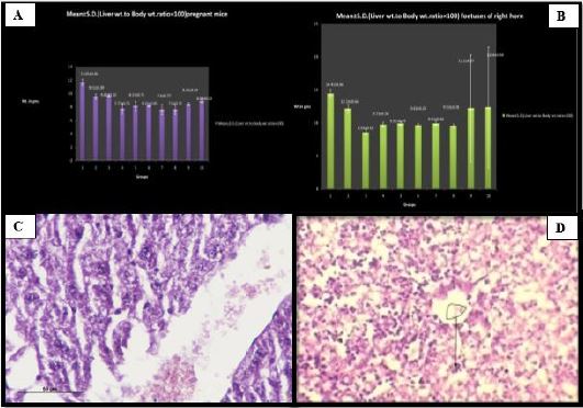
Apart from neurotoxicity these smallest, smaller medium and larger nano silver metallic particulate trap also causes vital organ toxicity and teratogenicity in mature and developing liver which is followed by dilatation and distortion of central vein, atrophy of hepatocytes in pregnant mice and loss of cord and rope with pile up pattern of developing hepatocytes with held of such particles in deep space results in multiple hemorrhagic focuses. The intensity of such histological alterations increases with increase in dose and size and amount of trap of nano silver metallic particulate.
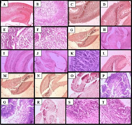
The control fetuses developing hippocampus histology (A & B) in H&E stain panoramic view (10 X & 40 X magnifications) shows absolutely normal CA1, CA2, CA3 & CA4 region with normal and aligned molecular, granular and pyramidal cell layers. Whereas in affected and treated developing hippocampus histology of AgNps colloidal solution treated Swiss Albino mice fetuses in chronological increase of dose and size manner (C-T= Group 2a, 2b, 2c; 3a, 3b, 3c; 4a, 4b, 4c) panoramic view shows distorted, degenerated and misaligned molecular, granular and pyramidal cell layer with mild, moderate to severe atrophy, disambiguation and degeneration of CA1, CA2, CA3 & CA4 region. The intensity of such periodically increases with increase of dose and size. (P≤ 0.004) (Group 2a towards Group 4c in fetuses group)
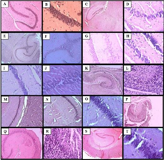
The control pregnant mice mature hippocampus histology (A & B) in H&E stain panoramic view (10 X & 40 X magnifications) shows absolutely normal CA1, CA2, CA3 & CA4 region with normal and aligned molecular, granular and pyramidal cell layers with adequate amount of cells in each layer. Whereas in affected and treated mature hippocampus histology of AgNps colloidal solution treated Swiss Albino pregnant mice in chronological increase of dose and size manner (C-T= Group 2a, 2b, 2c; 3a, 3b, 3c; 4a, 4b, 4c) panoramic view shows distorted, degenerated and misaligned molecular, granular and pyramidal cell layer with mild, moderate to severe atrophy, disambiguation and degeneration of CA1, CA2, CA3 & CA4 region and mild to severe loss of cells in each layer that is reduction in cell number . The intensity of such chronologically increases with increase of dose and size. (P≤ 0.001) (Group 2a towards Group 4c in pregnant mice group) Higher doses and larger size complications are grouped under hippocampus higher toxic or teratogenic effects.
Fast designing in animal AgNps treated modeling has led to advancements in understanding of fundamental disease mechanisms of many central nervous system disorders including silver nanoparticles trap in hippocampus tissue leads to initial cell death [48,49] motor and non motor ailment with axonal and dendrite degeneration and regeneration in traumatic brain injury by smaller to larger silver nanoparticles [50,51]. It is clear that a careful and meticulous study of animal modeling is essential as these models become incorporated into the translational process of neuropathic development and diagnostic virulent and lethal disease drug testing. Shoch et al. illustrate the power of serial molecular analysis with mouse transgenic applied to a pathologically complex event, such as traumatic injury by smaller and larger size nano metallic silver to mature and developing hippocampus that produces mechanistic results in studies of cell death and neuroplasticity. Finally leads to complex animal behavioral traits, such as addiction or defective behaviors, gene by gene by gene studies in mouse model are severely limiting [52].
We observed altered neurological and psychic states in pregnant mice and fetuses after repeated ingestion of admixture of smallest, smaller medium and larger size nano metallic silver at the time of giving medium and higher doses of AgNps colloidal solution. (P≤ 0.002) We observed anorexia nervosa, delirium, idiotic attitude in both pregnant mice and fetuses. We also observed fetuses’ sensed mothers’ psychological state while giving ingestion of AgNps colloidal solution as fetus grows, it constantly getting massage from its mother. It also gets chemical signals through the placenta. We also observed severe depression in mothers like sleeping silently in a corner of the cage in the end period of the dose. We also observed epileptic fits, festinate gate, focal seizures, convulsion in higher dose and larger size group in both pregnant mice and fetuses. (P≤ 0.003) So we came to a conclusion that traumatic hippocampus tissue injury causes altered neurological and psychic state in pregnant mice and their fetuses.
Discussion
In this present era nano metallic silver’s are vastly utilized in different clinical sub stream of medicine. It also
acts as best agent for drug vehicle device due to antiparasitic properties [1]. This type of nature resulted in
increase in homosapiens exposure ability. But the knowledge of the system wise toxicity and teratogenicity
show its relativity limited.
After going in vivo by injection nano metallic silver are spread in hippocampus tissue. Thus detail knowledge about the tissue illumination of such spread nano metallic silver is highly essential to understand the nature of smallest, smaller medium and larger size nano metallic silver inside the body. For identifying and calculate rate of risk, smooth illumination indicates a medium to low accumulative toxicity. Accordingly to it is also essential to enquire the concentration of nano metallic silver illumination from tissue following repeated oral gavages administration.
The objective of this study is to evaluate the rate of smallest, smaller medium and larger size with nine different dose (smallest, medium and larger) AgNps colloidal magnetic stirring cool solution and various size nano metallic silver illumination from mature and developing hippocampus tissue and its toxic and teratogenic ailments following repeated oral gavages administration (6 to 19 gestational days= 14 gestational days) in Pregnant Swiss Albino mice and their fetuses.
Three categories sizes of nano metallic silver. Smallest, smaller medium and larger was estimated by Uvvis spectroscopy, DLS & TEM exploration. Smallest indicate 0.27 to 1.03nm size range, smaller medium indicate 1.28 to 20.41nm size range and larger indicate 20.78 to 100.53nm size range. So a conclusion came out of this above procedure that this AgNps stirring fresh cool colloidal solution possesses an admixture of smallest, smaller medium and larger sizes with an avg. diameter of 71.9 to 300.1nm size ranges of nano metallic silver. Uv-vis spectroscopy and Dynamic Light Scaterring procedures also yeild the same result.
After this procedure the present ongoing research study clearly showed that AgNps used for repeated oral gavages experiment in Pregnant Swiss Albino mice and their fetuses can produce toxic and teratogenic sign and symptoms which are purely concentration, dose and size dependant manner. Section of developing and mature hippocampus and liver of pregnant Swiss Albino mice and their fetuses from control group showing normal and healthy histological structures in Fig. 7 (A, B) and Fig. 8 (A, B) while Fig. 6 (C-D) and Fig. 7 and 8 (C-T) showing histopathological section of mature and developing liver and developing and mature hippocampus treated with smallest, smaller medium and larger size with 9 different dose ( smallest, medium and bigger) nano (10-9 m.) metallic silver showing distraction and distortion with disambiguation in hippocampus and liver tissue in both. Gross edema or inflammation with honey comb vacuolation around the neuronal cell, hippocampus molecular, granular and pyramidal cells and hepatocyte cells are seen. The mature and developing liver histology showed centrilobular necrosis with abnormal dilatation of central vein that indicates the liver is the primary target organ for silver nano particle toxicity whereas hippocampus is the secondary because treated group shows dysmorphology, degeneration and distortion of molecular, granular and pyramidal cells with moderate to severe atrophy with misalignment and destruction in continuity tissue pattern of CA1, CA3 & CA4 region whereas CA2 destruction only observed in Nissel granule stain. The intensity of such sign and symptoms vary in 9 treated groups from lower towards the higher and it is size, dose and concentration dependant. Lower dose ailments are considered under toxic effects whereas moderate and higher dose are considered under teratogenic effects of hippocampus.
It is an established fact that silver ion and nano silver-based compounds are highly toxic on mother cells and animal cells. Because of to its tiny size it could penetrate into the blood vascular system by crossing the BBB and thus trap inside hippocampus tissue after traversing whole body but bigger size nano silver particles penetrate big vessels of body and big internal orifices of the body and cause multi vital organ and hippocampus teratogenic ailments after blocking macro and micro nutrition channels [6]. Thus nano silver particulate affect hippocampus and liver along with other vital organs such as lung, heart, Brain. In these organs nano silver cause teratogenic ailments such as cardiovascular diseases, pulmonary or lung swelling and hippocampus and neural teratogenicity [7]. The oral inject silver nano particles has a long absorption time and long tissue distribution inside the brain.
Cytotoxicity and neuroteratogenic effects are direct consequences of oxidative stress caused by medium and large size nano metallic silver and illumination of Ag ions. It strongly permeates cell membrane and plasma membrane by stimulating cell membrane hydrogen peroxide.
This research must be abide by many studies which shown that nano metallic silver alter and damage cellular processes by penetrating through cell membranes and tearing it due to small size and spiny and horny surfaces also it interact with intra cellular organelles leading to DNA and cell protein major damage. [5,8] and cross blood brain barrier to cause hippocampus and neurotoxicity [9]. It also disrupts BBB and induces brain inflammation. Therefore it is very important to know the various effects of smaller to larger size silver nano on hippocampus cell with special emphasis on possible mechanism and action. Larger and smaller size nano metallic silver may lead to inflammation of hippocampus tissue after chemically reacting with free radicals and Scavenger cells. In the year 2010 one nano research has demonstrated that larger and smaller size AgNps accumulate in rat brain micro vessel endothelial cell (rBMEC) in a size- dependant manner. It releases cytokine and other inflammatory mediators from the rBMEC cell single layer [10]. Still there is doubt these toxic or teratogenic ailments are due to ions illumination or itself nano silver metallic particles. Hippocampus toxicity and teratogenicity means both cytotoxicity and ion release. [11,12].
Conclusion
Maternal exposure to polyvinyl pyrollidone coated and sodium borohydried stabilized metallic nano silver
in colloidal form during pregnancy hampered balanced cognition and psychic state in pregnant mice and
offspring’s. Cognitive impairment purely dependant on one specific protein encoded gene depletion which
secretes from nerve ending and reduce neurite outgrowth. The hippocampus toxicity and teratogenicity was
mainly associated with disbursement of ionic silver.
Bibliography


Hi!
We're here to answer your questions!
Send us a message via Whatsapp, and we'll reply the moment we're available!