Biography
Interests
Andréa Barros Piazzon de Souza Queiroz, Laura Gusman Soares, Giovana Carvalho Vieira, Isabela Del Ponti, Alisson Diego Senna Fechis, Thiago André Salvitti de Sá Rocha & Fabrício Singaretti de Oliveira*
Department of Animal Morphology and Physiology, São Paulo State University (UNESP), Brazil
*Correspondence to: Dr. Fabrício Singaretti de Oliveira, Department of Animal Morphology and Physiology, São Paulo State University (UNESP), Brazil.
Copyright © 2019 Dr. Fabrício Singaretti de Oliveira, et al. This is an open access article distributed under the Creative Commons Attribution License, which permits unrestricted use, distribution, and reproduction in any medium, provided the original work is properly cited.
Abstract
Various are the alternative methods that look for the animal’s welfare in the veterinary surgery
teaching, which aim to substitute the use of live animals and causing similar or superior learning
to the students.
To determine the biomechanics effects depending on the intestine portions of fresh dogs’ corpses.
The maximum force and elongation of duodenum, jejunum and colon rupture were tested, and
compared among them so that future researches in biomechanics can know the resistance and
elasticity of those three intestinal regions.
Eight fresh dogs’ corpses were used and weighted 20.07±8.00Kg. Four samples were collected
of each portion of the intestine (duodenum, jejunum and colon) and immediately submitted to
biomechanical analysis.
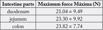
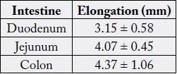
The force and elongation for duodenum, jejunum and colon rupture were 21.04±9.49N and
3.15±0.58mm, 23.30±9.92N and 4.07±0.45mm, 23.82±7.74N and 4.37±1.06mm, respectively.
Statistics indicated there was no difference among intestine portion as for maximum force (p=0.77)
and elongation (p=0.15) in rupture.
There was no significant difference in the duodenum, jejunum and colon resistance, but further
studies are needed so those dog cadavers used in Veterinary Medicine Course in surgery practicing
are the most similar to the fresh dog´s corpses.
Introduction
Duodenum, jejunum and ileum are portions of the small intestine and are responsible for finalizing food
digestion, nutrient absorption and endocrine secretion, and the large intestine (cecum, colon, rectum and
anal canal) is responsible for water absorption, mucus production and fecal mass formation [1]. Both have
simple columnar epithelium with striated edges and the goblet cells lie between the columnar cells. The
villi are restricted to the small intestine and they are long and thin, their base lies Lieberkuhn’s intestinal
glands or crypts, which are responsible for replacing the epithelial mucosa through cell division within the
crypt. The mucous muscle layer is formed by two layers of smooth muscle, separates the crypts from the
adjacent submucosa, and lies a sub-glandular lamina. The outer muscular layer is composed of smooth and
serous muscles that compose the rest of the intestine. In the initial and middle portions of the carnivorous
duodenum, the Brünner (duodenal glands and mucous glands), tubuloacinar and composite glands are
found [2].
In large intestine, the mucosa is smooth, without villi and crypts are long with abundant goblet and absorptive cells, but with a small number of enteroendocrine cells. The absorptive cells have short and irregular microvilli and are columnar. In carnivores, there are tubuloacinar anal glands in the submucosa and muscle layer and circum-anal glands around the anus [1].
There is an increasing interest on biomechanical properties of animal biological tissues. A great focus is given to comparative studies on tissues subjected to conservation and fresh tissues, generating data that contribute to the improvement of surgical techniques. Besides, there is a search for alternative biological material to be used as new option to animal experimentation [3].
Recent studies demonstrate biomechanics analyses of dogs and cats tissue during conservation [4-7], besides microbiological analysis of the fixative/conservative solutions [8] and students’ evaluation on surgery practice [9]. The objective of this paper was to determine, in fresh dog´s corpses, the maximum force and elongation in rupture of duodenum, jejunum, and colon, making statistical comparison among intestinal portions so that future researches in biomechanics can know the resistance and elasticity of those three intestinal regions.
Materials and Methods
Eight adult dog’s corpses weighing 20.07 ± 8.00Kg, male and female, were used and obtained from the
Zoonosis Center of Ribeirão Preto, São Paulo, Brazil, in a process approved by the Law Department (process
02.2014.000027-1). The animals were frozen (freezer at -18ºC) after death and then transported to the
Laboratory of Surgical Anatomy at the São Paulo State University (UNESP), Jaboticabal, São Paulo, Brazil,
located 50km away.
Corpses were thawed and shaved throughout the abdomen. The cadavers were placed on dorsal recumbency and a median abdominal incision with a scalpel was performed to expose the intestines. Duodenum, jejunum, and colon were identified and four samples of each were collected with a 1 x 5cm stainless steel mold [4]. Immediately after collecting, samples were subjected to biomechanical analysis.
To evaluate tissue resistance, a Universal Testing Machine (EMIC® - DL 2000) was used, with a 50N load cell and electromechanical drive support, in a speed of 100mm/min. Traction claws were also used by manual compression, in the Laboratory of Surgical Anatomy of the Department of Animal Morphology and Physiology of São Paulo State University (UNESP), Jaboticabal, Brazil.
The Shapiro-Wilk and Kolmogorov-Smirnov were used to verify the data normality, and then they were submitted to Kruskal-Wallis test to make the statistics.
Results
Mean and standard deviation of the maximum force and elongation for duodenum, jejunum and colon
rupture of the fresh dogs are shown on table 1 and 2.
Data from the maximum force and elongation were subjected to Shapiro-Wilk and Kolmogorov-Smirnov tests (P< 0.05) and the distribution was nonparametric. The Kruskal-Wallis test indicated no significant difference among duodenum, jejunum and colon as for the maximum force (p=0.77) and elongation (p=0.15).
Discussions
The use of live animals in research and teaching activities has changed and we must seek alternatives to
their use mainly in surgery practice. Recently, in Brazil, 75.67% of the students from a Veterinary College
approved the use of chemically preserved cadavers (with ethylic alcohol in fixation and sodium chloride
solution in tanks for conservation) in the teaching of surgery and 81.08% were in favor of the initial surgical
training on cadavers, followed by practice on animals submitted for elective surgery at a Veterinary Hospital
[9]. In another study of Silva et al, 2003, there was 93.29% of acceptance in favor of this teaching method
with chemically prepared corpses. In chemically prepared cats (with curing salt and ethylic alcohol), statistics
did not point difference among scores when compared to fresh corpses in incision (p=0.8055) or even in
suture (p=0.5022) [4].
Besides, chemically prepared dogs (with ethylic alcohol in fixation) were tested, using a surgical microscope, in suture of the external jugular vein (score 4.0 of 5.0) [6] or common carotid artery (score 4.5 of 5.0) [7]. After conservation (in tanks with sodium chloride solution), scores were 3.5 of 5.0 for both.
The force necessary to cause rupture on duodenum (21.04N), jejunum (23.30N) and colon (23.82N) of dogs weighting 20.07±8.00Kg in this research was lower to the force to rupture jejunum of dogs weighting 7.6±2.7Kg (27.6N) [5] maybe because the stainless steel mold was different.
Intestines are very soft tissues, what would explain why the necessary force for rupture was much lower than those to cause rupture in skin of fresh dogs (131.3± 75.6N) [5] or fresh cats (254.15±183.25N) [4]. However, that force was similar to the one necessary to rupture the fresh external jugular vein (19.98±11.49N) [6] or the common carotid artery (25.77 ± 15.43) of fresh dogs [7].
Conclusions
There was no significant difference in the duodenum, jejunum and colon resistance, what is very satisfactory
on researches using those intestinal portions to biomechanical analyses.
Bibliography


Hi!
We're here to answer your questions!
Send us a message via Whatsapp, and we'll reply the moment we're available!